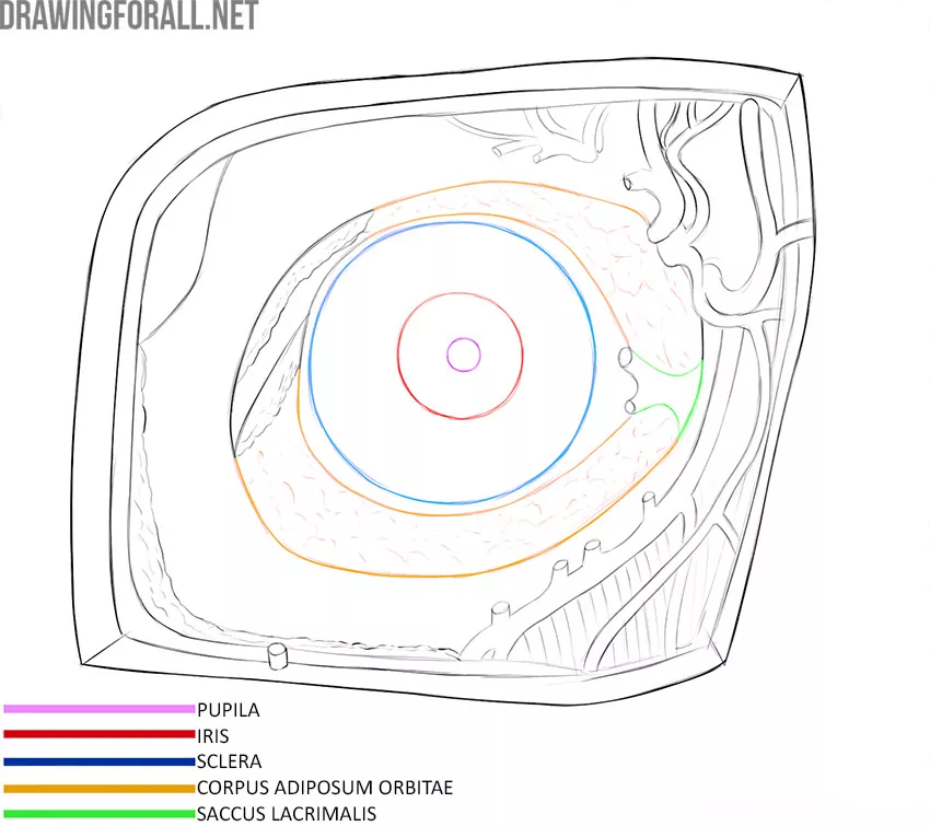
Anatomy of the Eye
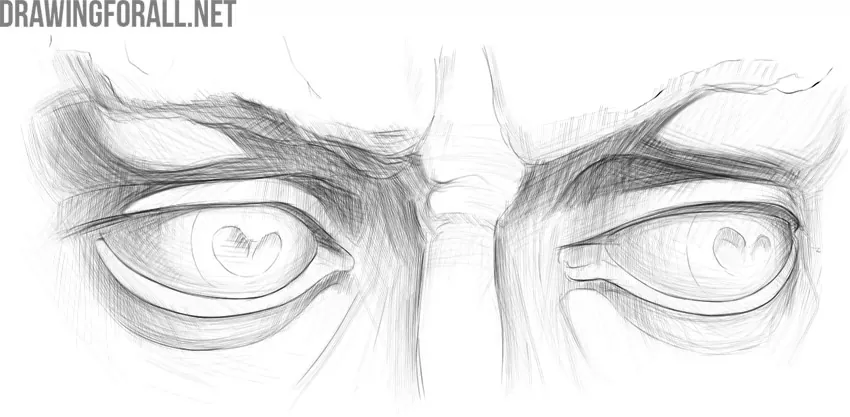
The eyes are one of the most important parts of the human face. The eyes reflect the character’s emotions and mood. In many styles of drawing, eyes are the main focus. You can see this effect in anime style, for example.
Today we will tell you about the anatomy of the eye. More precisely, about those aspects of eye anatomy that are important for artists.
Some basic knowledge of anatomy is the same for artists, doctors, or scientists. First of all, it is necessary to understand that the eye consists of the eyeball and the auxiliary apparatus of the eye. The auxiliary apparatus of the eye includes the eyelids, lacrimal apparatus, eyebrows, and eye muscles.
Eyeball Anatomy
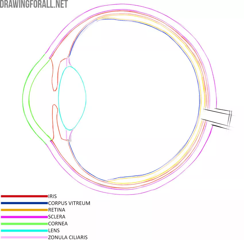
The eyeball consists of three membranes that cover the nucleus of the eye. Nothing fancy, just three shells and a core – it’s pretty easy to remember, isn’t it? Now we will talk a little about each of the three shells.
Fibrous Membrane
This shell is the most superficial. The fibrous membrane of the eye is quite dense, and it forms a rigid frame for all other parts of the eyeball.
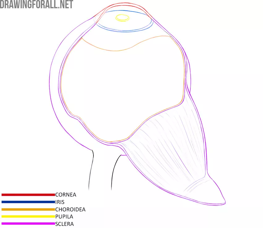
The fibrous membrane consists of two parts – the most protruding part of the fibrous membrane is called the cornea. This is one of the most transparent areas of the eyeball. The cornea must be completely transparent because it is the first to encounter a beam of light that must travel through the entire eye.
The second part of the fibrous membrane is called the sclera and is a completely opaque layer. The opacity of the sclera is very important so that light is not scattered on its way through the eye.
Choroid
The choroid is the middle layer of the eyeball. For artists, the fundamental point is that it is this shell that forms the color of the eye. The choroid is rich in pigments melanin, and the color of the eye depends on the amount of melanin. A large amount of melanin corresponds to brown eyes, a little less to green eyes. The minimum amount of melanin is found in the choroid of a person with blue eyes.
The choroid has an opening called the pupil. Contrary to popular belief, the pupil is not an independent structure of the eye; the pupil is the opening through which we can see the deepest layer of the eye.
Retina
It is this shell that is the main structure of the entire eye. The task of all other shells and light-refracting mediums is to somehow deliver the light reflected from objects to the retina. The retina contains receptors that pick up light rays and convert them into nerve signals.
The nucleus of the Eye
The nucleus of the eye contains several light-refraction mediums, that is, substances through which light passes before it reaches the retina.
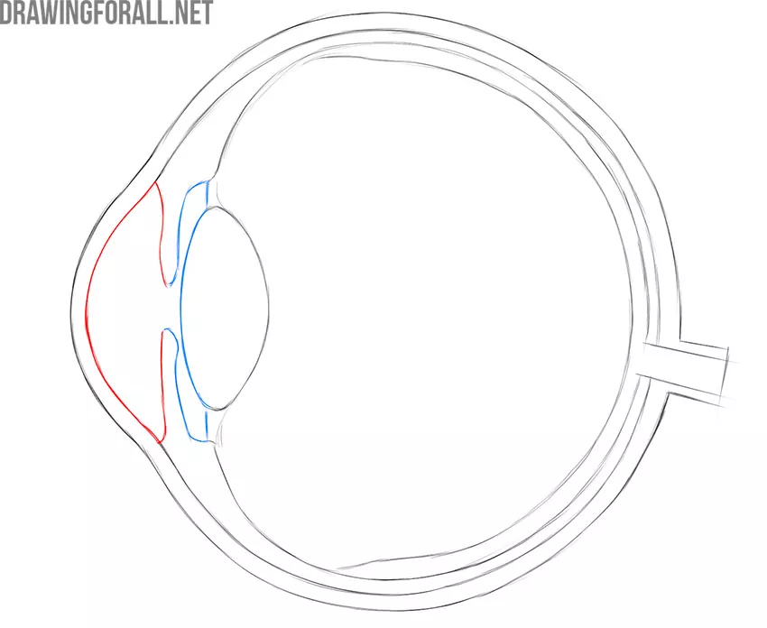
Anterior Chamber
The anterior chamber is the space between the cornea and the iris that is constantly bathed by a small amount of intraocular fluid called aqueous humor. Aqueous humor in a healthy person is absolutely transparent.
Lens
The lens is a small, flexible lens made of a completely transparent substance. The lens is attached to ligaments that connect to the ciliary muscles. Muscle contraction changes the shape of the lens, which is why we can see objects close and very distant from us with the same quality.
Posterior Chamber
The posterior chamber is a small slit-like space between the iris and the muscles that are filled with clear fluid. This is the same fluid that washes the anterior chamber.
In this illustration, the anterior chamber of the eye is highlighted in red and the posterior chamber of the eye is highlighted in blue.
Vitreous
It is the largest part of the eye that forms the rounded shape of this organ. The vitreous has a gel-like consistency. The vitreous humor, like all other light-refracting mediums, is completely transparent.
Accessory Organs of the Eye
The accessory organs of the eye are anatomical structures that help the eye to perceive visual information as efficiently as possible. In this article, we will only cover those structures that are important for plastic anatomy.
Eyebrows
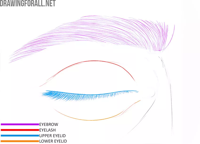
Eyebrows are small bumps located above the eye sockets and are covered with short, coarse hair. Brows are the natural protection for the eyes from sweat and excess light.
Eyebrows can have a variety of shapes. Some people have a very light eyebrow color and it may seem to you that there are no eyebrows at all. Older men may have very thick, dark, or gray eyebrows. The position of the eyebrows helps convey various emotions.
Eyelids
The eyelids are movable areas of skin that are located above and below the eyes. The eyelids protect the eyes from dust, sweat, and other annoyances, and they also completely insulate the eyes if necessary.
The eyelids are also a way to convey emotion. Wide-open eyes show strong emotions such as joy or deafening rage. Half-closed eyelids are typical for a calm and phlegmatic mood.
Lacrimal Apparatus
The lacrimal apparatus is a complex of tissues and organs that control the production and removal of tear fluid. We cannot see many parts of the lacrimal apparatus on a living person, however, we can see the lacrimal lake and the lacrimal meat in the medial corner of the eye. Both of these formations help to drain excess tear fluid.