Skull Anatomy
The skull is the skeleton of the head. We can also say that the skull is an extremely important bone structure that protects everything that helps us live and exchange information with the world. The skull forms reliable protection for the brain, organs of vision, hearing, smell, touch, and taste. It is also necessary to remember about the pyramidal tracts, the control of vital centers, and the glands. In general, the skull is a very useful thing.
And today the team of Drawingforall.net will tell you the basic anatomy of the skull in order to make it easier for you to draw a skull, and also to better represent the bone base of the human head.
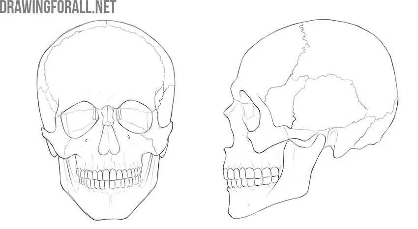
So, the human skull consists of 23 bones. The human skull is divided into two major sections – the cranium and the facial skeleton.
Cranium
The cranium forms the cavity of the skull in which the cerebrum is located.
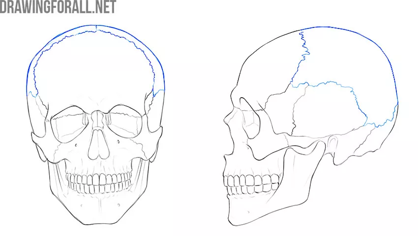
The cranium is composed of bones that we can see from the outside of the skull and bones that we can only see on the skull with a sawn top. The frontal, parietal, temporal, and occipital bones are those bones that we can see on the skull.
To see the sphenoid and ethmoid bones, we need to remove off the top of the skull.
Frontal Bone
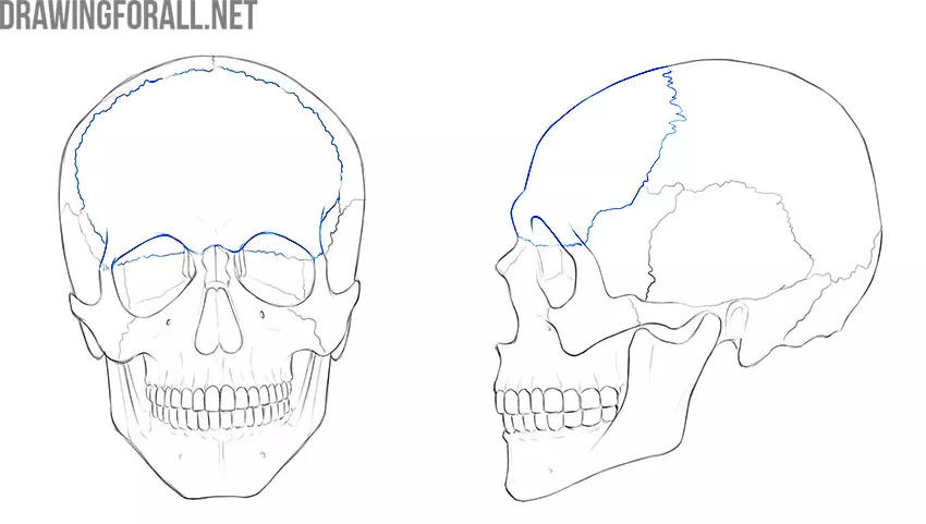
The frontal bone is located in the front, above the eyes and nose. The frontal bone connects with the parietal bones from above and with the temporal bones laterally.
The lower edge of the frontal bone forms the upper walls of the orbits. Also, the center of the lower edge forms the upper part of the nose. The frontal bone has convex sections called frontal tubercles. Below them, you can see the arched protrusions called the superciliary arches.
The lower edge of the frontal bone forms the upper walls of the orbits. Also, the center of the lower edge forms the upper part of the nose. The frontal bone has convex sections called frontal tubercles. Below them, you can see the arched protrusions called the superciliary arches.
The frontal bone plays a huge role in shaping the image of a person. Small mistakes in drawing the superciliary arches or incorrect calculation of the height of the frontal bone can distort even the best portrait beyond recognition.
Parietal Bones
These are paired bones. Thus, there are right and left parietal bones that form the upper part of the top of the skull. The parietal bones connect with the frontal bone in front, with the occipital bone in the back and with the temporal bones laterally.
The parietal bone has four edges. The squamous edge is the junction with the temporal bone. The squamous edge has a clearly visible concave bend inward. The occipital margin is the junction with the occipital bone, you can see a smooth convex bend here. The sagittal edge is the junction with the other parietal bone, usually a flat edge with minimal bending. The frontal margin is located in front and adjacent directly to the frontal bone.
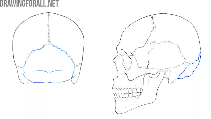
Occipital Bone
If we remove the top of the skull, we will see that the cavity of the skull looks like three pits (fossa) – front, middle, and back.
The posterior cranial fossa is formed by the occipital bone. It is in the posterior cranial fossa that the vital centers of the brain are located that are responsible for respiration and vascular tone. Damage to these centers is extremely dangerous for life, therefore the occipital bone is a very important bone.
The occipital bone connects in the front with two parietal bones and laterally with the temporal bones. Also on the lower outer surface of the occipital bone, you can see protruding flat tubercles – these are places for connection with the first cervical vertebra. At the bottom of the occipital bone is a large opening, called the large occipital opening. It is through this hole that the part of the brain that connects it to the spinal cord passes.
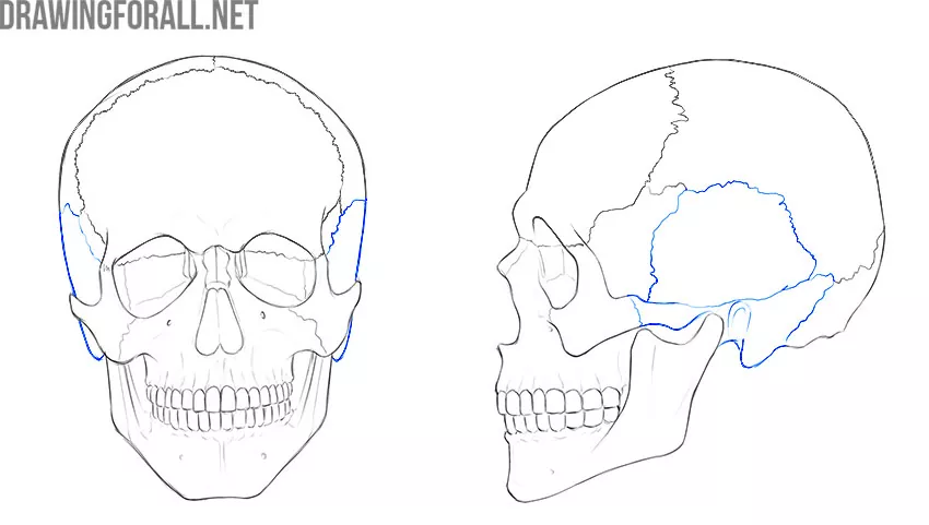
Temporal Bone
The temporal bone connects to the occipital bone in the back, the parietal bone from above, and also with the sphenoid bone in the front. The temporal bone also connects to one of the bones of the facial skull, namely the lower jaw.
The temporal bone has a rather complex internal structure.
The temporal bone has two clearly visible parts – the pyramid and the squamous part, as well as the tympanic part, which does not matter in plastic anatomy. In the thickness of the pyramid are the channels of the temporal bone, in which the most important vessels and nerves pass, such as, for example, the internal carotid artery and facial nerve.
However, the channels of the temporal bone are also not important for plastic anatomy.
The temporal bone has several clearly visible processes that form the appearance of the skull. First of all, it is the zygomatic process, which is directed towards the facial skull and connects to the zygomatic bone. You can also see and even feel the rounded mastoid process, which is located directly under the ear. There is also the styloid process that is covered with soft tissues and does not create visible contours on a living person.
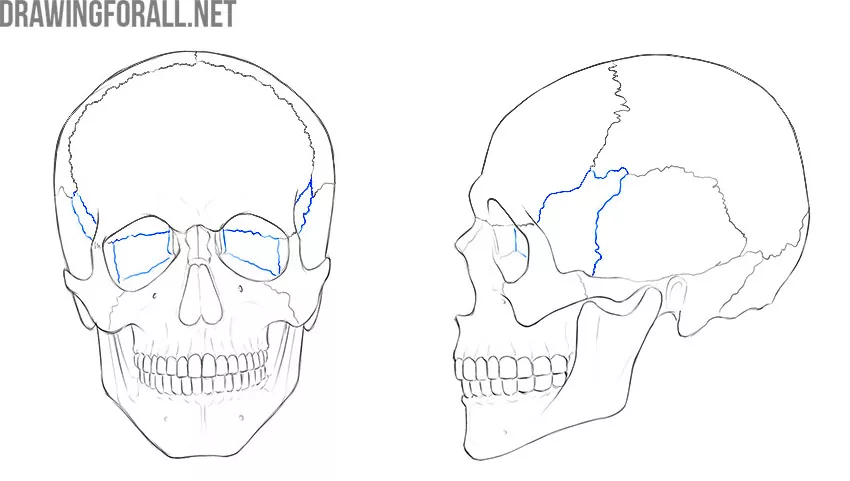
Sphenoid Bone
Many artists forget about the sphenoid bone because when we look at a whole (un-sawn) skull, we can see only small parts of this bone.
The sphenoid bone has a corpus and two large flat processes, which are called the large wings of the sphenoid bone. The sphenoid bone also has small wings, which are located slightly above the large wings.
We can see parts of the large wings if we look at the skull from the side. The area that will be visible to us from this angle on the whole skull is called the temporal surface of the large wing of the sphenoid bone. We can also see small parts of large wings if we look at the skull from the front. The posterior wall of the orbit is formed partially by the large wing of the sphenoid bone. This part is called the orbital surface of the large wing of the sphenoid bone.
In the corpus of the sphenoid bone is located a very important bone deepening. This bone, called the sella turcica is a small cozy hole in which the pituitary gland is located – the most important endocrine gland.
Ethmoid Bone
The ethmoid bone is located in the anterior cranial fossa. This small bone is pierced by numerous openings through which vessels and nerves pass – mainly the fibers of the olfactory nerves. The ethmoid bone does not have much significance in plastic anatomy, because it cannot be seen on the whole (un-sawn) skull.
Facial Bones
The facial section of the skull forms several important holes at once. The eye socket is a reliable wall for the organ of vision, the oral cavity is the beginning of the digestive system, and the nose is the first echelon of air purification that enters our lungs. Let’s see which bones form the basis for these anatomical structures.
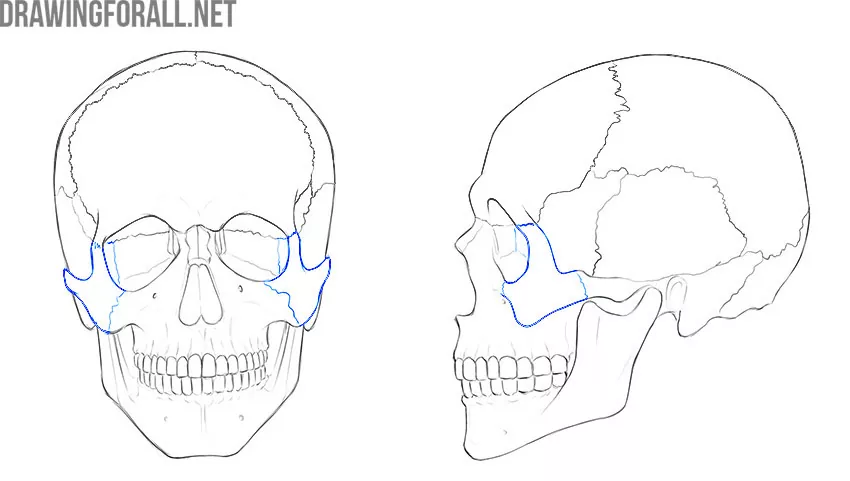
Zygomatic Bones
The zygomatic bone is located laterally and below the organ of vision. The zygomatic bone forms the lower and lateral wall of the orbit. Also, the shape of the face, especially in thin people, significantly depends on the shape of the zygomatic bone.
Each zygomatic bone has two clearly visible processes that connect this bone with three other bones of the skull:
⦁ The frontal process is located on top. This process connects the zygomatic bone with the frontal bone;
⦁ The temporal process is directed back and laterally to connect to the zygomatic process of the temporal bone.
The zygomatic bone also connects to the upper jaw. A fairly long section of this joint is called the infraorbital suture.
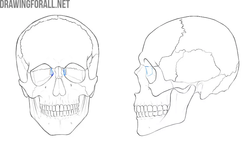
Lacrimal Bone
This small bone is clearly visible if we look into the eye socket of a well-prepared skull. The lacrimal bone looks like a small quadrangular plate that lies in the medial wall. The upper edge of this plate connects to the orbital part of the frontal bone, and the lower edge connects to the upper jaw.
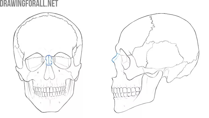
Nasal Bone
These are small, but rather important bones. For some reason, many textbooks on plastic anatomy do not mention nasal bones, which is rather strange.
The right and left nasal bones are located directly under the conditional center of the frontal bone. The nasal bones form the upper part of the bone base of the nasal cavity. You can easily palpate the nasal bone if you touch the wings of your nose and move slightly towards the frontal bone.
The upper edge of the nasal bone is connected with the frontal bone, the lateral one – with the upper jaw. Also, the inner surface of the nasal bone is connected to the plate of the ethmoid bone, however, we do not see this connection on the skull.
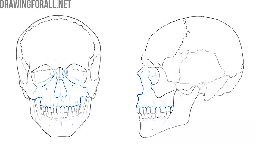
Upper Jaw
Contrary to the existing stereotype, the upper jaw is not one solid bone. There are two upper jaws that connect in the center of the face. So, the upper jaw is a large paired bone that forms a significant area of the face from the orbits to the oral cavity.
Each upper jaw has a body and four processes. One of these four processes we can not see on the whole skull, and the rest are clearly visible and accessible for palpation:
⦁ Frontal process. This is the highest located process of the upper jaw, its upper edge is connected to the frontal bone;
⦁ Zygomatic process. With this process, the upper jaw connects to the zygomatic bone;
⦁ Alveolar bone. This is the lower surface of the upper jaw, in which there are holes for the roots of the teeth;
⦁ Palatine process. It is this process that is hidden from our eyes. But then your dentist can palpate it because it forms the upper wall of the oral cavity;
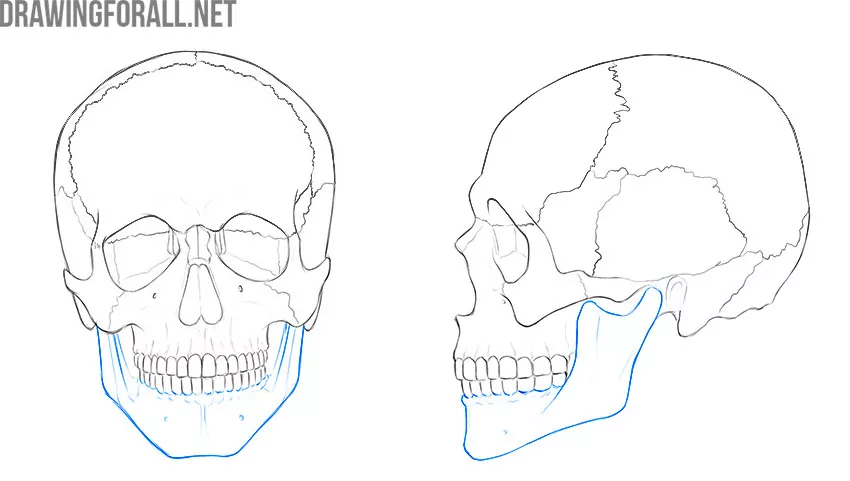
Lower Jaw (mandible)
Unlike the upper jaw, the mandible is one solid bone. The lower jaw is quite remarkable. Among all the bones of the skull, only the lower jaw and temporal bone form a full-fledged, mobile joint.
The lower jaw is also very important in plastic anatomy because the shape and expression of the face largely depend on the shape and position of the lower jaw.
The mandible has a corpus and two branches – the right and left. The transition of the corpus of the mandible to the branch is called the angle of the lower jaw. This angle is dull and fairly smooth, rounded. Each branch ends with two large processes – the condylar process and the coronoid process. The condylar process is the place of the formation of the temporomandibular joint. The condylar process is the place of attachment of the powerful temporal muscle.
In older people, the body and branches of the lower jaw tend to thin out – this is due to the gradual loss of teeth and a decrease in the load on bone tissue. In this case, the characteristic position of the lower jaw is formed, when it is noticeably pushed forward.
Inferior Nasal Concha, Vomer, and Palatine
These bones are involved in the formation of the nasal and oral cavities. We cannot see them without a preliminary cut of the skull in the sagittal plane, therefore, in plastic anatomy, they are not of fundamental importance.
It is very important for the artist to know human anatomy in order to accurately draw portraits, making them voluminous and realistic. The information from this article, along with knowledge about perspective, proportions, light and shadow, is the basis for any artist. If you want to learn how to create compelling and realistic drawings, then be sure to study the articles from the Basic Drawing Lessons category on Drawingforall.net. And also do not forget to subscribe to Drawingforall.net on Twitter, Facebook, and other social networks to keep abreast of new articles on our website.


