Neck Bones Anatomy
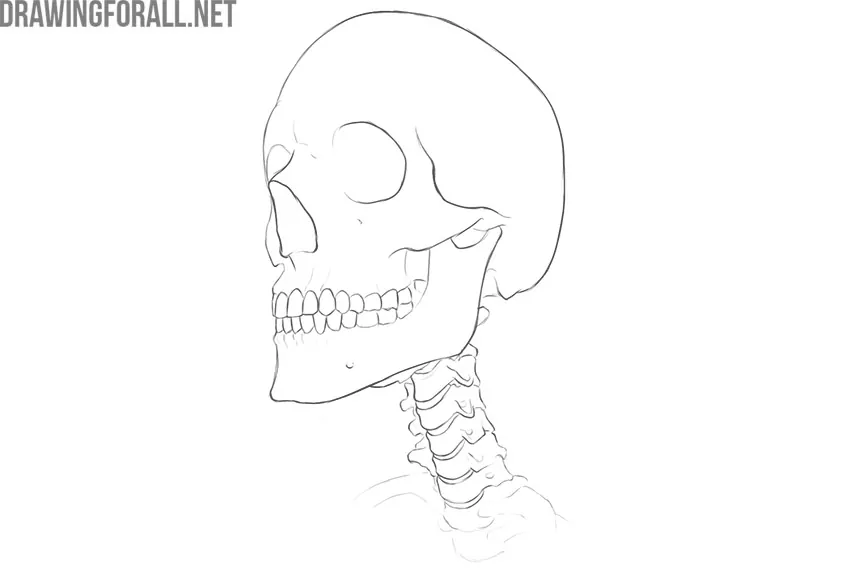
The neck is a very interesting and unusual part of the body in anatomical terms. The neck includes vital organs and blood vessels and does not have any bone protection. The only bones we can see when examining the neck are the vertebrae that form the cervical spine. And today we’ll talk about it.
The vertebral column is an unusually important bone structure that is the axis of support for the entire human body. The vertebral column was formed precisely in the course of millions of years of evolution. A huge influence on the structure of the spinal column was made by upright posture and the need to lean on two legs, holding the trunk in an upright position.
In addition to the function of support, the spinal column also performs the function of protecting the spinal cord. That is why there is a hole in the body of each vertebra. In several vertebrae standing on top of each other, these holes form the spinal canal. In this canal, the spinal cord is located, with the help of which we carry out movements and have the ability to experience sensations, such as touch, pain, a sharp increase in temperature, and others. That is why injuries to the spinal column are always very dangerous.
The vertebral column is represented by vertebrae – short dense bones that are interspersed with intervertebral discs. The intervertebral discs consist of elastic cartilage and soften the shock load during jumping, running, and other dangerous actions for the spine. Also, the spinal column is additionally strengthened by powerful ligaments and muscles.
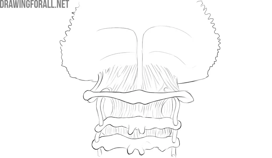
The vertebral column is divided into several parts:
⦁ Cervical;
⦁ Thoracic;
⦁ Lumbar;
⦁ Sacrum;
⦁ Coccyx.
The vertebrae of each part differ in shape and size. Moreover, each part is formed by a different number of vertebrae.
Typical Cervical Vertebrae
An ordinary cervical vertebra is small in comparison with more massive thoracic and lumbar vertebrae.
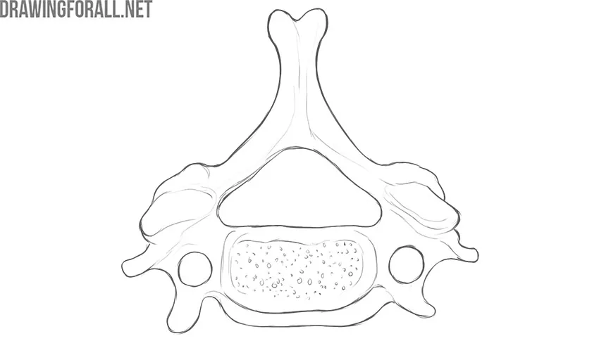
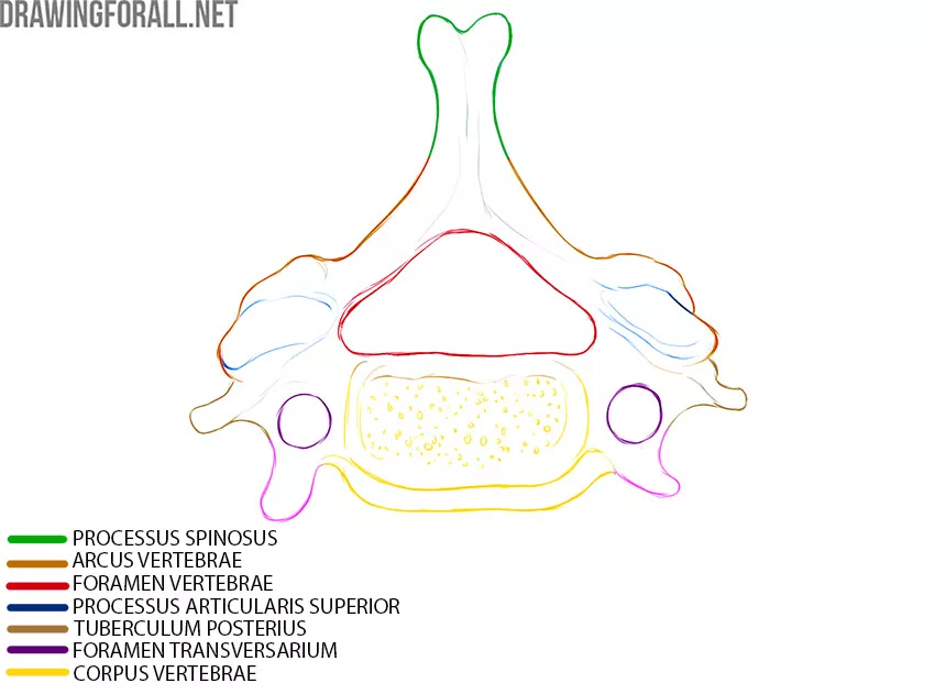
If you look at the vertebra, you will immediately see a dense bone mass – this is the body of the vertebra (corpus vertebrae). The main support from the weight of the vertically located head and from the shock load on the lower extremities falls on the vertebral body. Shock-absorbing intervertebral discs are located precisely on the vertebral corpus above and below.
In the back of the body of the spine is the vertebral arch (arcus vertebrae). The vertebral body and the vertebral arch form the vertebral foramen (foramen vertebrae), which we have already talked about. The spinal cord located in these holes.
Lateral to the arc of the vertebra are the transverse processes (processus transversus). The transverse processes of the cervical vertebrae are very unusual, and you can easily identify the cervical vertebra in several ways:
⦁ Small size
⦁ Front and back tubercles
⦁ The presence of a transverse hole (foramen transversarium)
The cervical vertebrae also have articular processes. In fact, the articular processes are in the vertebrae of all parts.
Atlas
Atlas is the first cervical vertebra. Its structure is significantly different from all other cervical vertebrae.
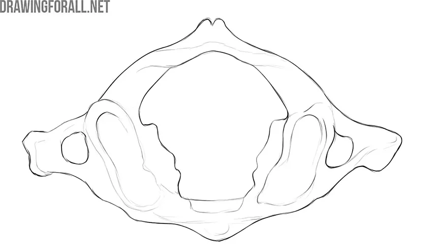
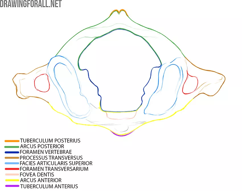
First, the atlas does not have a body. Instead of the body, we can see the front (arcus anterior) and back (arcus posterior) arches that connect and form the vertebral foramen (foramen vertebrae).
The thickenings that are at the junction of the anterior and posterior arches are called lateral masses. On the upper and lower sides of the lateral masses are articular surfaces for articulation (facies articularis) with the skull and the second cervical vertebra, respectively.
On the inside of the front arc of the atlas, we see a small fossa called the fossa of the dens (fovea dentis).
Axis
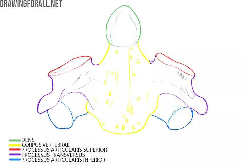
Axis is the second cervical vertebra. The first and second vertebrae form an atlanto-axial joint that rotates the head in different directions.
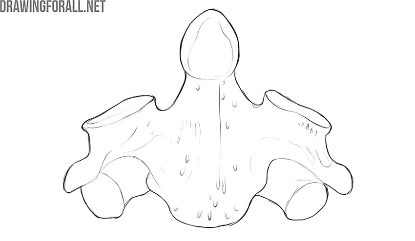
Axis, or the second cervical vertebra, has a clearly visible vertical process – the dens. The rounded upper edge of this process is called the tip of the dens. The dens articulate with the fossa of the tooth, which is located on the anterior arch of the first cervical vertebra. It is this joint that allows us to turn our heads around.
Both first cervical vertebrae are very small and light in weight. Their anatomical features are determined by rather specific functions, which are associated, first of all, withholding the skull in an upright position and performing various head movements.