Lower Limbs Skeleton Anatomy
The skeleton of the lower limbs and the vertical spinal column is a unique evolutionary device that allowed a person to raise his head above all other living creatures on our planet.
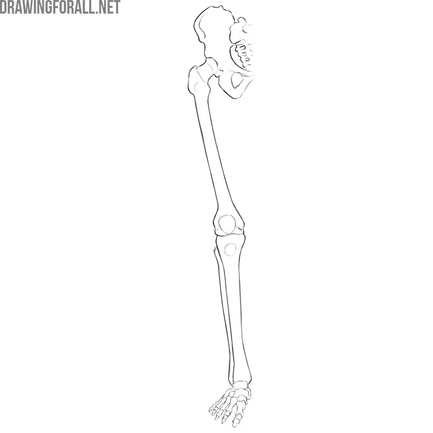
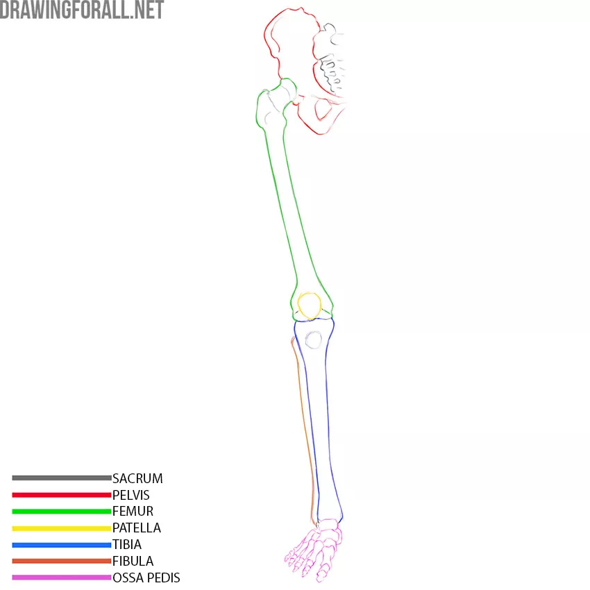
The lower limb skeleton includes the cingulum membri inferioris (the lower limbs bones belt) and skeleton membri inferioris liberi (that is the legs). The cingulum membri inferioris is the bones of the pelvis that connect to the sacrum.
The lower limbs are divided into three parts – thigh (femur), lower leg (tibia and fibula), and foot. Bony main foot are ossa pedis.
So let’s take a look at each component of the lower limb skeleton.
Lower Limbs Bones Belt
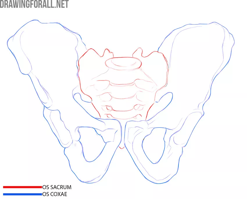
The lower limb belt consists of two pelvic bones (os coxae) – the right and left, which are connected to the sacrum. Each of the pelvic bones consists of three more bones (looks like a nesting doll, doesn’t it?).
The pelvic bones are of great physiological importance. The pelvic bones form the cavity of the small pelvis, in which the vital organs, great vessels, and large nerve trunks that innervate the entire lower limb are located. Also, the pelvic bones are involved in the process of childbirth. It is very important for obstetricians and gynecologists to correctly measure the distance between certain points of the pelvic bones of pregnant women in order to predict problems that may arise during childbirth.
In addition, the pelvis, as you already know, is the girdle of the lower limbs. This means that the pelvic bones serve as a support for the lower limbs and a link between the lower limbs and the spinal column.
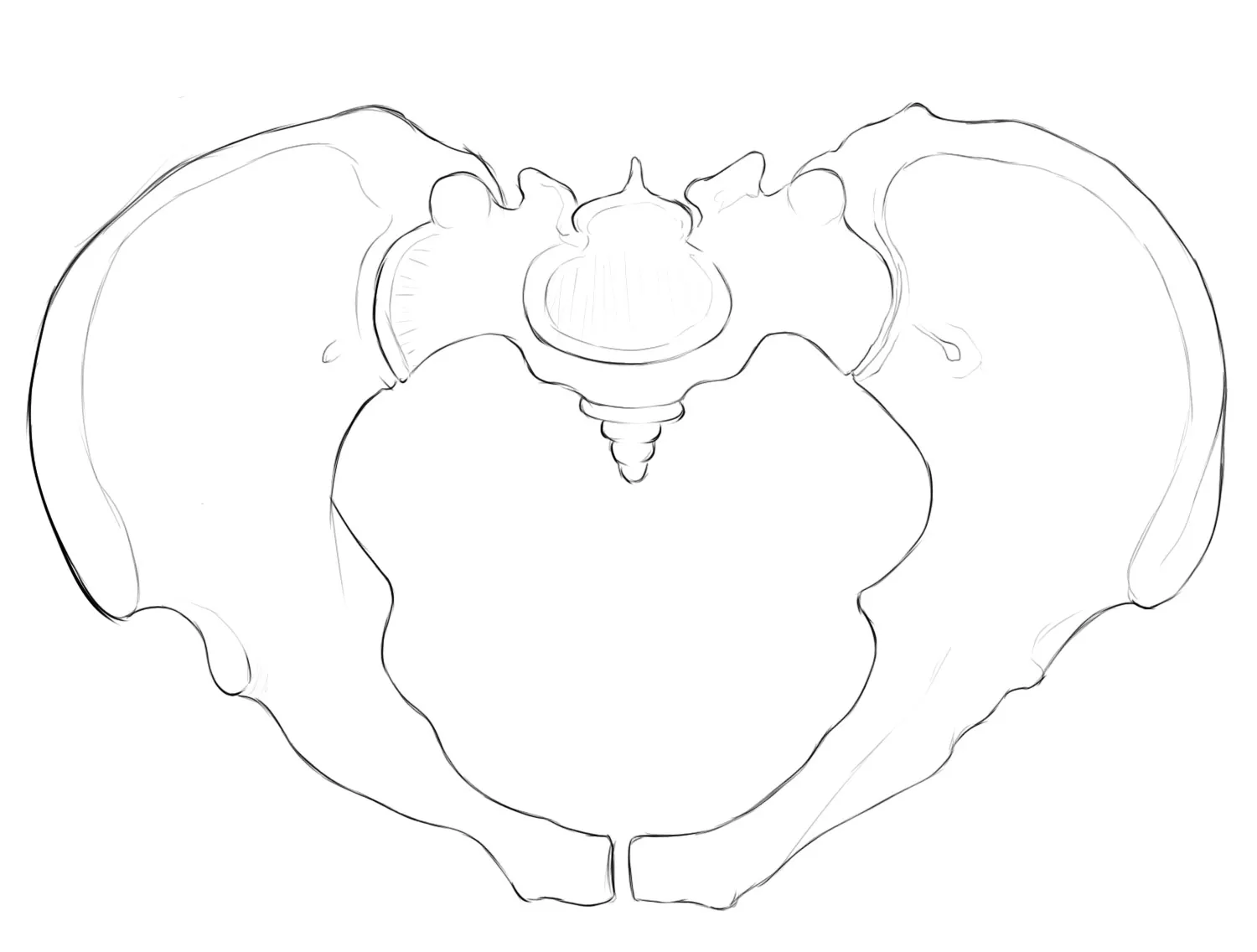
So, the pelvic bone consists of the ilium, ischium, and pubic bones. These three bones are connected by cartilage in childhood, and in adolescents, they grow together into a single bone.
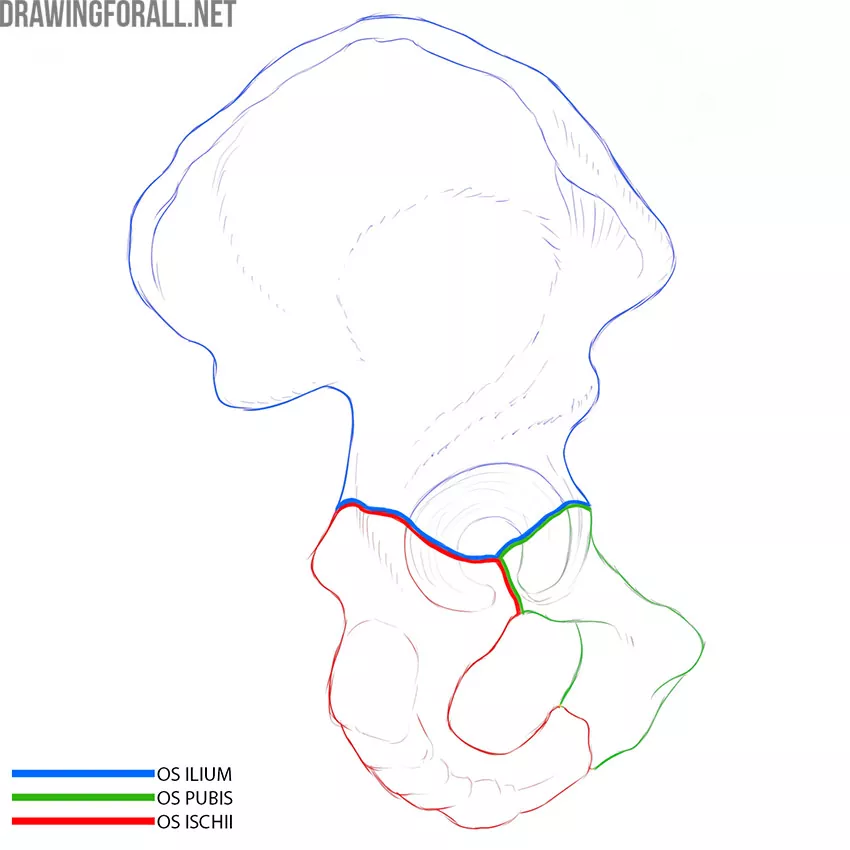
Each bone has several component parts. For example, the ilium has a body and a wing. The ischium has a body and one branch, and the pubis has a body and two branches. As you can see, each bone has a body – a small, dense, rounded area.
The bodies of the three bones grow together, forming the acetabulum – the junction of the pelvic bone and the rounded head of the femur. This forms a highly mobile and multifunctional hip joint.
The female pelvis has several differences from the male pelvis. We will focus on two of the most notable ones:
- The junction of the two pelvic bones in front is called the pubic symphysis. In men, the pubic symphysis forms an acute angle, and in women, an obtuse angle;
- The wings of the ilium in men are vertical and narrow. In women, the wings of the ilium open wider and the pelvis becomes visually wider.
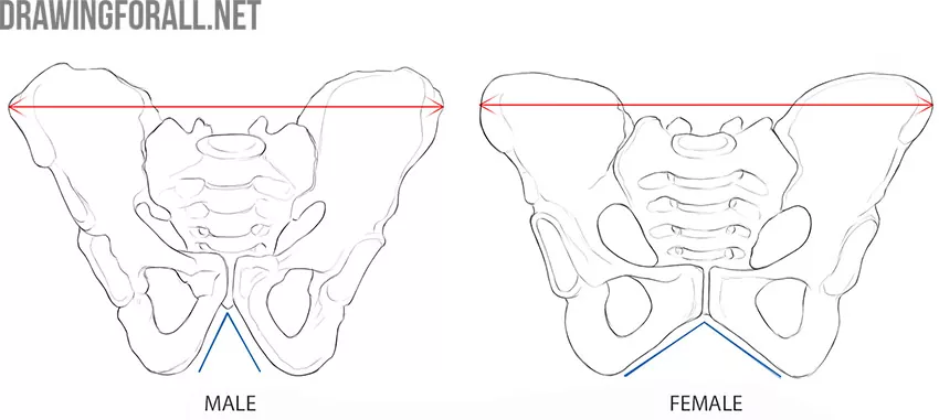
It is always very important to determine the characteristics of the figure of the person you are drawing. The female pelvis is always wider and larger. This is especially noticeable in relation to the width of the shoulders – a typical male figure is characterized by wide shoulders and a narrow pelvis, and the female is on the contrary.
Femur
The femur (or the thigh bone) is one of the most massive and strong bones in the body. The femur is articulated with the pelvic bone at the proximal end and with the shin (tibia) at the distal end. There is a large rounded protrusion at the proximal end of the femur, it is the head of the femur (caput femoris). This shape allows for maximum mobility of the hip joint.
The connection of the femur to the pelvis has a different shape in men and women. If you look at the men’s skeletone, you can see the head and body of the femur form an angle close to the right angle. An obtuse angle is more typical of the female skeleton. This affects the contours of women’s hips – it is because of this feature that women’s hips look wider and rounder.
The head of the femur goes into a narrower part called the neck of the femur (collum femoris). You can also see a large bony protrusion that is located laterally. This is the greater trochanter (trochanter major), where the thigh muscles attach. And there is another small protrusion called the lesser trochanter (trochanter minor) more medially. It is also where the thigh muscles attach.
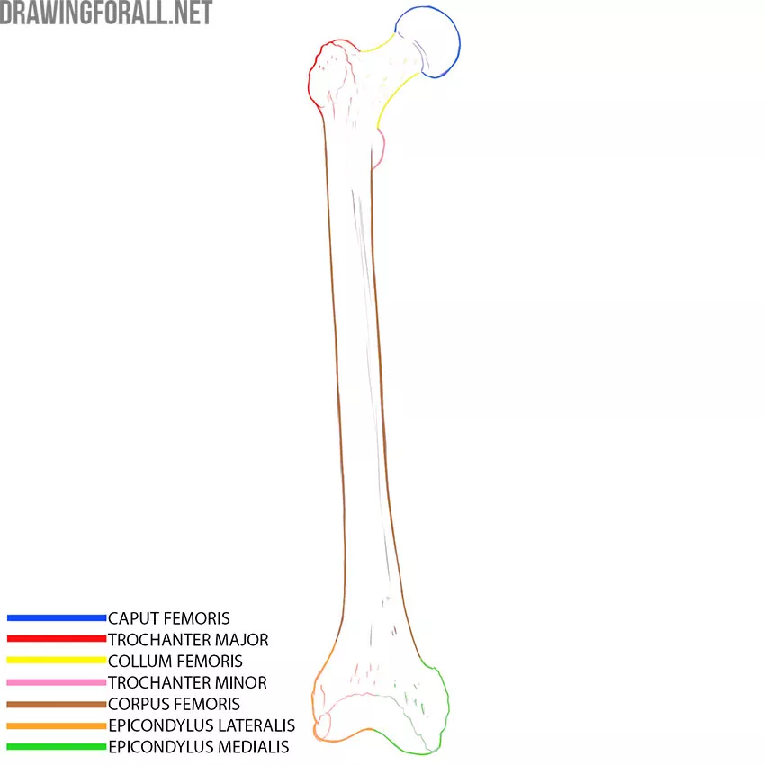
The main part of the femur is called the femur body (caput femoris). Downward, the body expands and forms medial epicondyle (epicondylus medialis) and lateral epicondyle (epicondylus lateralis). These are small elevations that are located above the articular surfaces of the femur. The articular surfaces themselves are called condyles. We can see it if we look at the femur from below.
Patella
The patella is a small but dense bone that is part of the bones of the knee joint. The patella is fixed by the tendons of the thigh muscles, it covers the knee joint and makes it more stable, preventing unnecessary movements in the medial and lateral directions.
The patella also connects to the powerful cruciate ligaments that provide additional stability to the knee joint.
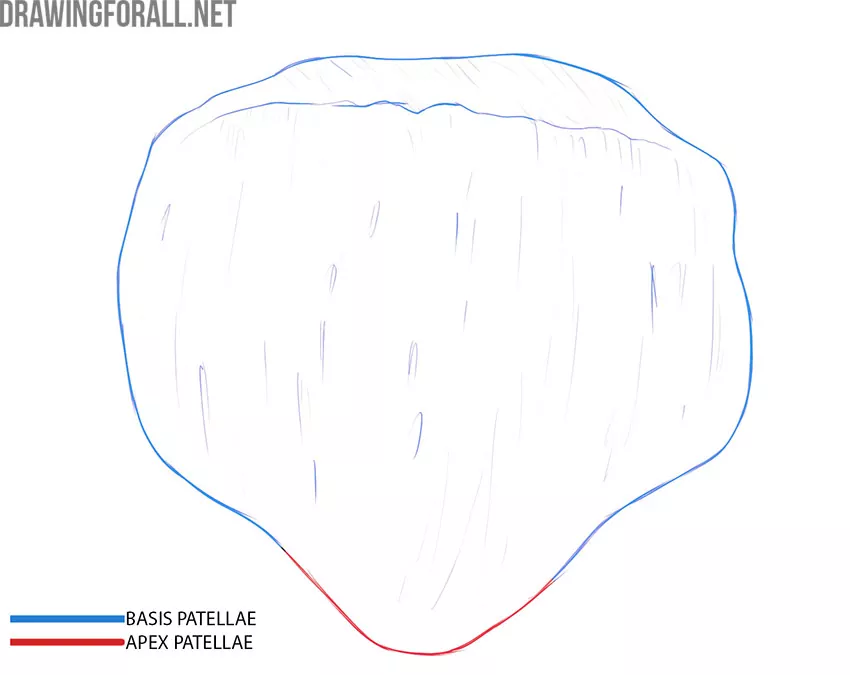
The patella has a base, an apex, and two surfaces. This small bone looks like a flat stone with a distinct edge on one side.
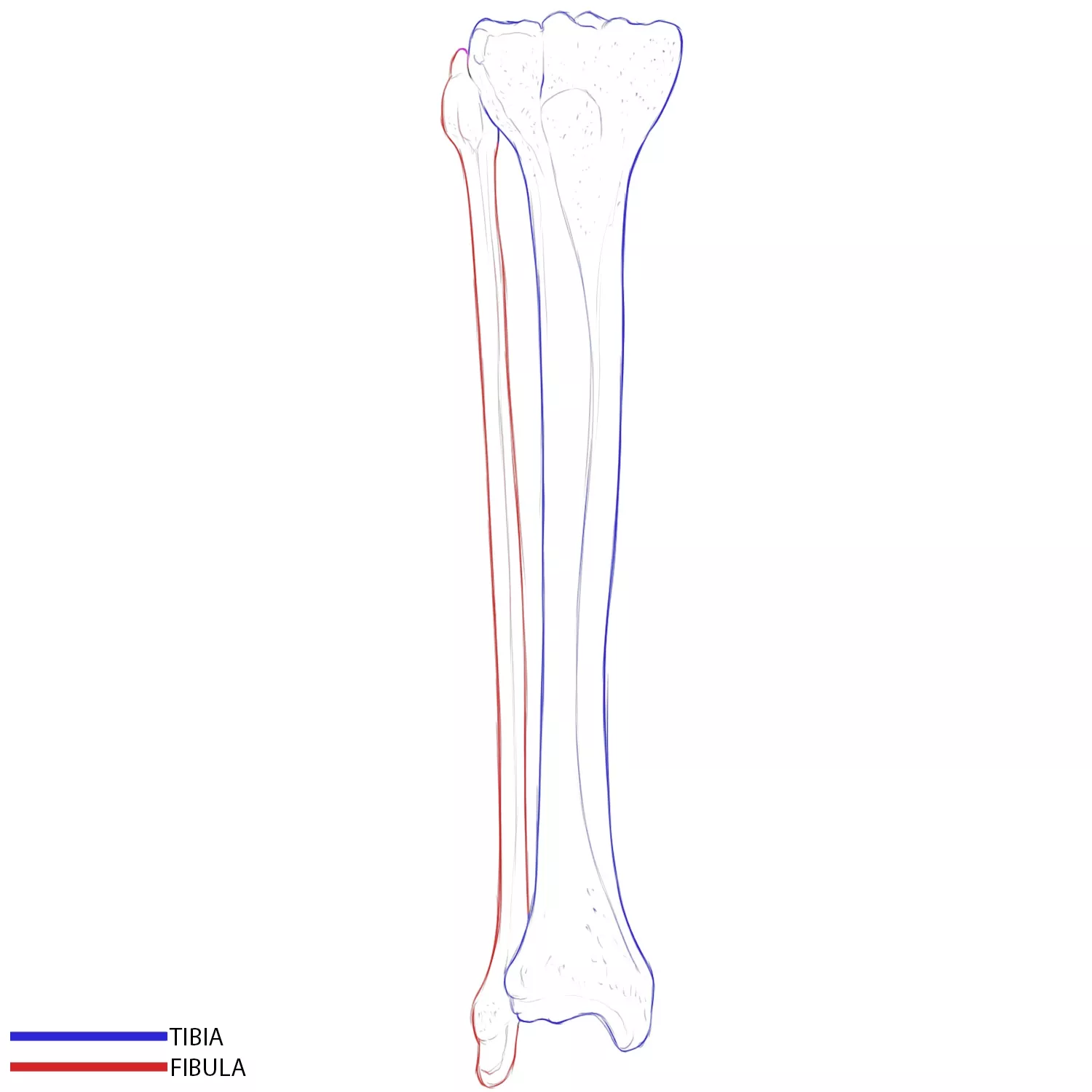
Tibia
The tibia is a very massive, strong bone that connects to four bones at once. The proximal end of the tibia articulates with the femur, the distal end with the talus of the foot. On the lateral side, the fibula adjoins the tibia.
Like all tubular bones, the tibia has a body (corpus tibiae) and two ends – the upper and lower. The powerful body of the tibia expands upward to form two large condyles to connect to the femur (condylus lateralis/ condylus medialis).
Downward, the body of the tibia expands less noticeably. Here we can see a small rounded protrusion on one side and a flat surface on the other side. The rounded protrusion is the medial malleolus, and the flat surface is the fovea for articulation with the fibula. On the medial side of the area, which is highlighted in pink, is the incisura fibularis.
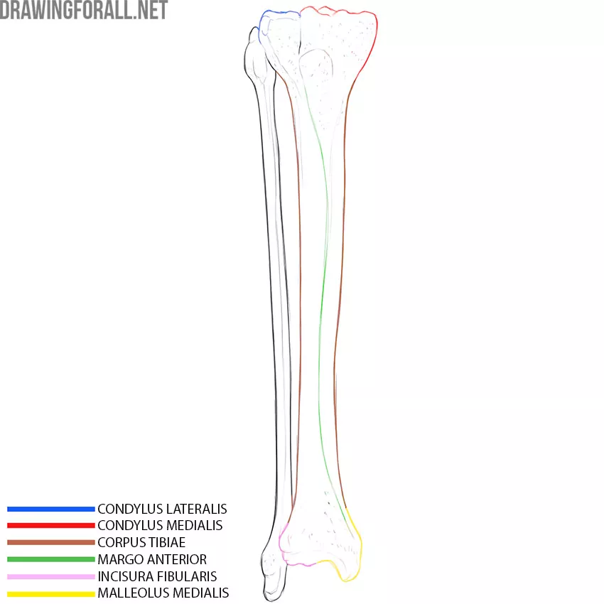
The tibia has a trihedral form for most of its length. The front edge of the tibia (margo anterior) is contoured through the skin. You can also palpate it easily.
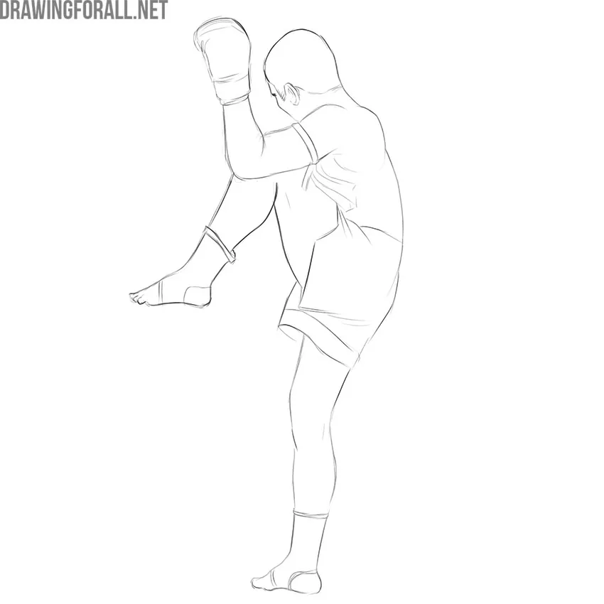
By the way, Muay Thai fighters (a popular form of the strike martial arts) use the shin as a shield, protecting themselves from hits and exposing the hard front edge of the tibia to attack.
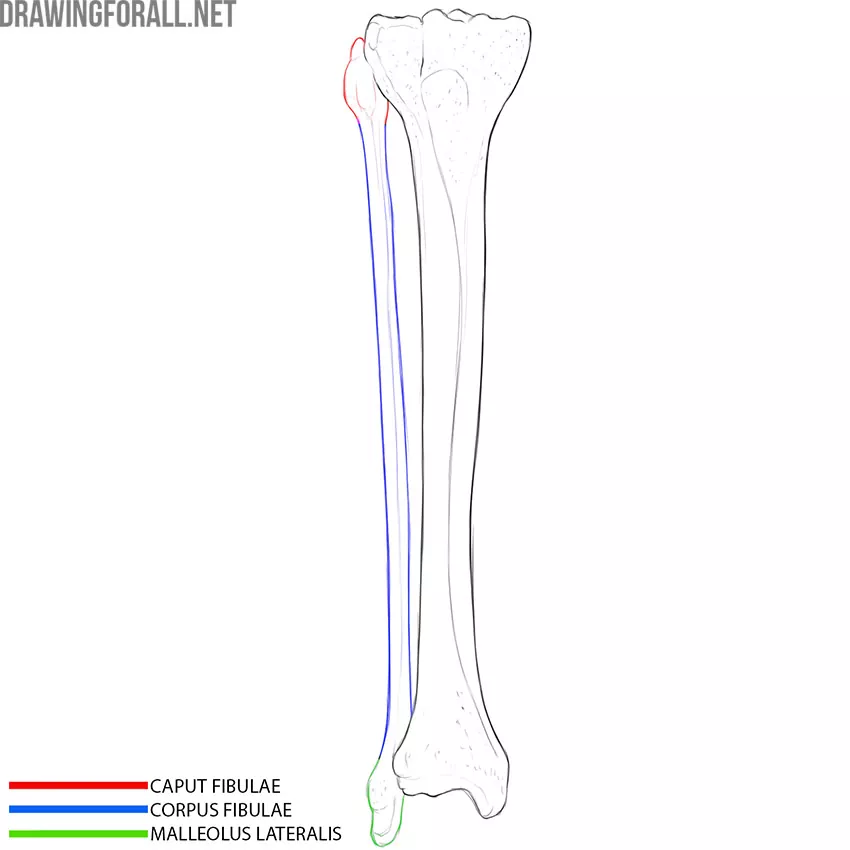
Fibula
I think that the structure of the fibula will not be a surprise to you because this bone has a body, an upper end, and a lower end. The body of the fibula is much less massive than that of the tibia.
The proximal end of the fibula is slightly rounded outside – this is the head of the fibula. Inside this end, there is a small depression for articulation with the tibia.
The lower end of the fibula forms the lateral malleolus, which looks like an elongated bump. If you look at the fibula, you might think the lateral is meant to be articulated with the tibia. In fact, the lateral malleolus articulates with the talus of the foot. The articulation with the tibia is much higher.
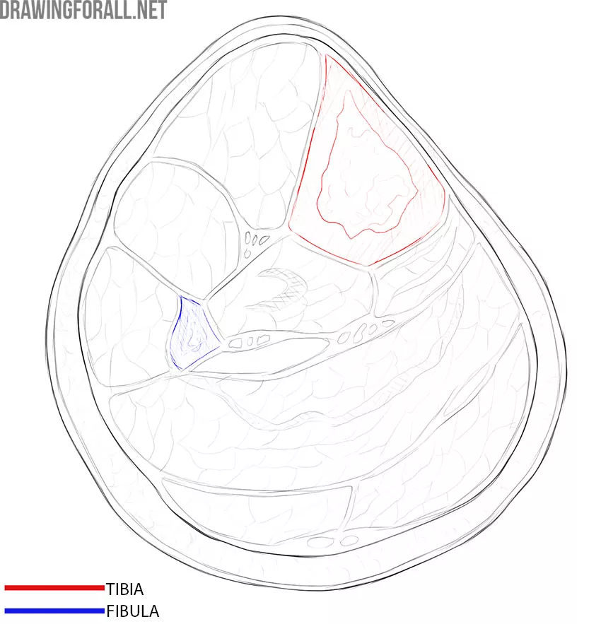
A dense connective tissue membrane is located between the tibia and the fibula. In addition to this membrane, many muscles are located between the bones, as well as in front and behind. This is how the tibia and fibula look on a horizontal cut in the mid-shin:
Foot Bones
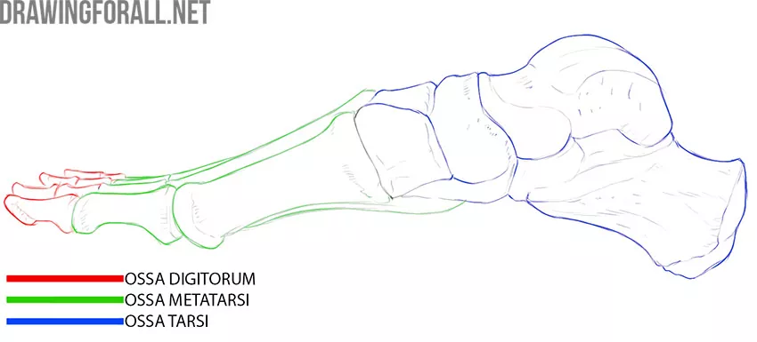
The human foot is a unique evolutionary device designed for running, jumping, and walking on two legs. The foot consists of the bones of the tarsus, metatarsus, and phalanges of the toes.
The tarsus bones include two rows of bones. The proximal row includes the talus and calcaneus. The distal row includes the scaphoid, cuboid, and three wedge-shaped bones.
The talus and calcaneus are very strong and dense bones. The talus is grasped from above by two tibial bones, forming the ankle joint. The tibia wrap around the medial and lateral ankle surfaces in a fork-like manner.
In addition to the two tibias, the talus articulates with the calcaneus, as well as with the scaphoid and cuboid bones.
If you look at the calcaneus, you immediately see a large bump that goes into a slight curve. This construction, reinforced by muscles and ligaments, allows us to jump from a sufficiently high height and run at high speed without injuring the bones of the upper limb or spine.
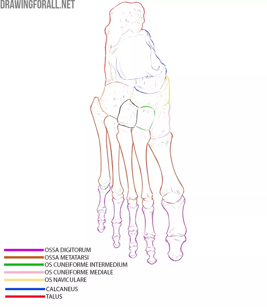
If we go further distal, we see five bones of the metatarsus and bones of the toes. The bones of the toes are called phalanges. Each toe, except for the first, consists of three phalanges – distal, proximal, and middle. The big toe has only the distal and proximal phalanges.
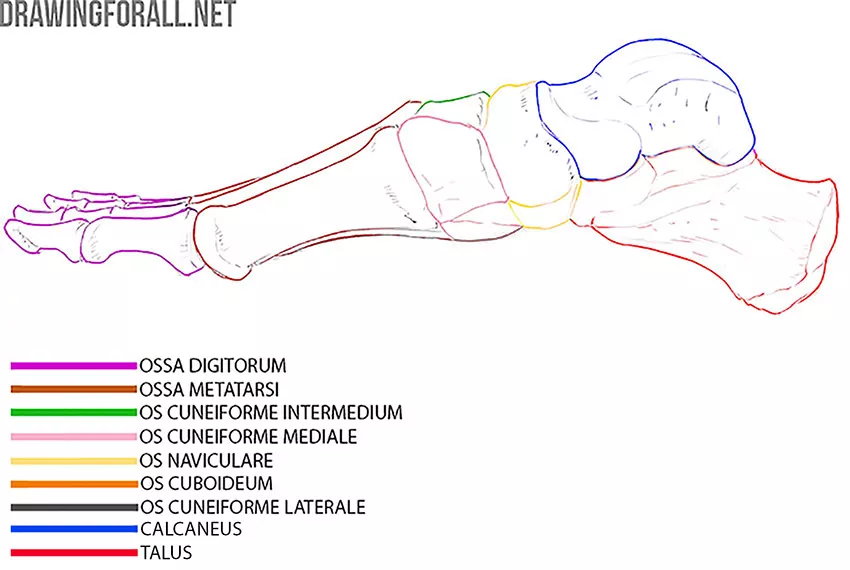
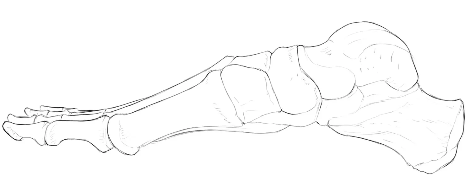
The metatarsal bones are very important. It is they that form the characteristic bend of the foot, which is also called the arch. This bend is necessary to cushion jumps and other shock loads.
This bend is especially noticeable when we look at the foot in profile:
The bones of the metatarsus, like the phalanges of the toes, do not have special names, they are simply numbered in the direction from the metatarsus of the big toe to the metatarsus of the little toe


