Head Muscles Anatomy

The muscles of the head are divided into two large groups – the muscles of facial expression and the muscles of mastication.
The main functions of the muscles of facial expression are to convey emotions through facial expressions and to participate in protective reactions such as blinking or sneezing. The main function of the muscles of mastication is to move the lower jaw to grind food.
The muscles of facial expression have some distinctive features. As you know, bones are the attachment points for most skeletal muscles. A muscle attaches to bones, usually near a joint. When contracting, the muscle acts on the bones, sets the joint in motion, and as a result, we see movement.
Unlike most other skeletal muscles, many muscles of facial expression attach to the skin of the face and move it when they contract.
Another feature of many facial muscles is that they are not enclosed by deep fascia in the same way as most skeletal muscles. Fasciae are layers of connective tissue that surround and separate muscles and other anatomical structures.
Finally, one more feature concerns the topography of the muscles of facial expression. Many of these muscles are located around the natural openings of the face, such as the eyes, nose, and mouth.
Let’s look at the main muscles of the face and head that artists should know to build a solid foundation in drawing.
Epicranius Muscle
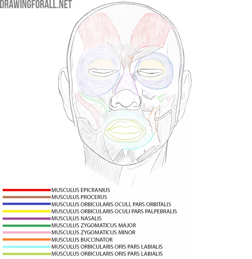
The epicranius is a broad muscle of facial expression. It has two main parts – the frontal belly and the occipital belly. Both parts are connected by a dense tendon sheet called the epicranial aponeurosis, or galea aponeurotica.
The frontal belly extends from the epicranial aponeurosis to the skin of the eyebrows and forehead. The occipital belly extends from the superior nuchal line of the skull to the posterior part of the epicranial aponeurosis.
The occipital belly pulls the scalp backward, while the frontal belly raises the eyebrows and wrinkles the skin of the forehead.
Orbicularis Oculi
It is a large, prominent muscle that surrounds the eye. The orbicularis oculi is divided into three parts: the orbital part, the palpebral part, and the lacrimal part.
The orbital part is located around the orbit. This is the largest part of the muscle. When it contracts, it closes the eyelids tightly and wrinkles the skin around the eye.
The palpebral part forms the muscular layer of the eyelids. Its contraction gently closes the eyes.
The lacrimal part is very small and difficult to see without anatomical dissection. It assists in the drainage of tear fluid.
Procerus Muscle
It is a small muscle located between the eyes over the upper part of the nose. When it contracts, the procerus pulls the medial parts of the eyebrows downward and creates transverse folds over the bridge of the nose.
Nasalis Muscle
The nasalis muscle has two parts. The transverse part extends across the side and bridge of the nose and passes into an aponeurosis over the dorsum of the nose.
The alar part lies around the wing of the nose and attaches to the alar cartilage.
The two parts have different actions. The transverse part compresses the nasal opening, while the alar part helps widen the nostril.
Orbicularis Oris Muscle
Like many muscles of facial expression, the orbicularis oris has several groups of fibers. It includes labial and marginal parts. The labial part lies within the lips, while the marginal part surrounds it.
When contracted, the orbicularis oris closes the lips and can also protrude them forward.
Buccinator Muscle
The buccinator is a broad muscle that forms much of the muscular wall of the cheek. It lies deep to several other facial muscles and is covered superficially by the buccal fat pad, which is especially prominent in infants.
When both buccinator muscles contract, they press the cheeks against the teeth and help keep food between the teeth during chewing. The muscle can also help pull the corner of the mouth laterally.
Zygomaticus Major and Zygomaticus Minor
These two muscles are fairly easy to find because they both begin from the zygomatic bone. They have a similar direction and are located close to each other. To distinguish them, remember that the smaller zygomaticus minor lies closer to the nose.
The zygomaticus major extends to the corner of the mouth, while the zygomaticus minor blends with the upper lip.
Both muscles participate in facial expressions. The zygomaticus major pulls the corner of the mouth upward and laterally, as in smiling. The zygomaticus minor raises the upper lip and helps deepen the nasolabial fold.
Muscles of Mastication
The muscles of mastication are large, powerful, and essential for chewing. A person has four pairs of masticatory muscles, of which two are of particular importance for plastic anatomy because they contribute to the visible contours of the face.
Temporalis Muscle
The temporalis muscle is a large, flat muscle that fills the temporal fossa. Unlike the other muscles of mastication, the temporalis is located on the side of the cranial vault. It begins from the temporal fossa and the temporal fascia.
Its muscle fibers converge downward, forming a strong tendon that attaches mainly to the coronoid process of the mandible.
You can feel the contraction of the temporalis muscle if you place your fingers on the temporal region and clench your teeth several times.
Masseter Muscle
This is also a powerful and highly visible muscle. The masseter becomes clearly visible through the skin when it contracts, especially in lean people.
The masseter begins from the zygomatic arch and attaches to the lateral surface of the ramus and angle of the mandible. Its main function is to elevate the mandible during chewing.

Basic Topography of the Head
In topographic anatomy, body regions are studied rather than individual organ systems. Here we will look at the basic topography of the head. This includes not only the skull but also the muscles and other anatomical structures that form important landmarks for plastic anatomy.
Head Borders
The border between the head and neck runs from the chin along the lower edge of the mandible to the mastoid process, then along the superior nuchal line to the external occipital protuberance.

Cerebral and Facial Parts of the Head
The skull forms the skeleton of the head, so the boundaries of its facial and cerebral parts correspond closely to the facial and cerebral parts of the head.
The boundary between the cerebral and facial parts of the head can be traced from the brow region toward the zygomatic arch and then toward the upper edge of the external auditory opening.

Head Regions
Determining the boundaries of regions is an important task in topographic anatomy. To study the regions of the head, we must be able to distinguish clearly between its cerebral and facial parts.
Regions of the Cerebral Part of the Head
On the cerebral part of the head, we can distinguish three main regions – the frontoparieto-occipital region, the temporal regions, and the mastoid regions. The frontoparieto-occipital region is unpaired, while the temporal and mastoid regions are paired.
The frontoparieto-occipital region is bordered:
⦁ In front – by the supraorbital margins of the frontal bone;
⦁ Behind – by the superior nuchal line;
⦁ Laterally – by the superior temporal lines.
The temporal region is bordered:
⦁ In front – by the zygomatic process of the frontal bone and the frontal process of the zygomatic bone;
⦁ Behind – by the posterior boundary of the temporal region;
⦁ Above – by the superior temporal line;
⦁ Below – by the zygomatic arch.
Mastoid Region
The mastoid region corresponds to the area over the mastoid process.
Regions of the Facial Part
Orbital Region
The boundaries of the orbital region correspond approximately to the margins of the bony orbit:
⦁ Above – the supraorbital margin of the frontal bone;
⦁ Below – the infraorbital margin formed by the maxilla and zygomatic bone;
⦁ Laterally – the zygomatic bone;
⦁ Medially – the frontal process of the maxilla and the nasal part of the frontal bone.
Nose Region
The boundaries of the nasal region correspond to the contours of the external nose, including the root, dorsum, apex, and wings.
Zygomatic Region
The boundaries of the zygomatic region correspond approximately to the contours of the zygomatic bone.
Infraorbital Region
The infraorbital region borders:
⦁ Laterally – the zygomatic region;
⦁ Medially – the nasal region;
⦁ Above – the orbital region;
⦁ Below – the buccal region.
Buccal Region
The buccal region borders:
⦁ Medially – the nasolabial fold and oral region;
⦁ Laterally – the parotid-masseteric region;
⦁ Below – the lower edge of the mandible;
⦁ Above – the zygomatic and infraorbital regions.
Mouth Region
The oral region is bordered:
⦁ Above – by the nasal region;
⦁ Below – by the chin region;
⦁ Laterally on both sides – by the buccal regions.
Chin Region
The chin region is bordered:
⦁ Above – by the oral region;
⦁ Laterally on both sides – by the buccal regions;
⦁ Below – by the lower edge of the mandible.
Parotid-Masseteric Region
The parotid-masseteric region borders:
⦁ Above – the zygomatic arch and temporal region;
⦁ Below – the lower edge of the mandible;
⦁ Medially – the buccal region;
⦁ Posteriorly – the region of the auricle.

With this basic knowledge of the head muscles, it will be much easier to draw the human face by understanding and constructing its form rather than simply copying lines.