Upper Limbs Muscle Anatomy
The upper limb is not just a hand. As you already know, in addition to the skeleton of the shoulder, forearm, and hand, the scapula and clavicle are also referred to the bones of the upper limb. The scapula and clavicle are the girdle bones of the upper limb. Thus, there are bones of the free upper limb and bones of the girdle of the upper limb.
This principle is also observed in myology. There are the muscles of the free upper limb, that is, the arms, and the muscles of the girdle of the upper limb, that is, the muscles associated with the shoulder blade or clavicle.
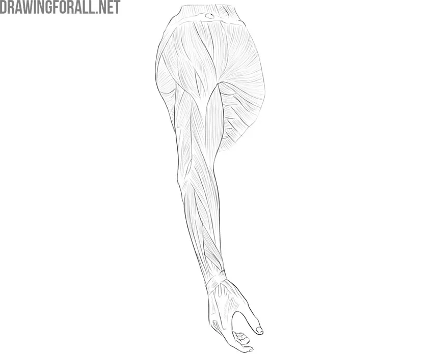
As you know, we present you with the basic aspects of the human body that are important to artists. Therefore, we will only tell you the large superficial muscles that form the appearance of a person.
Muscles of the Upper Limbs Girdle
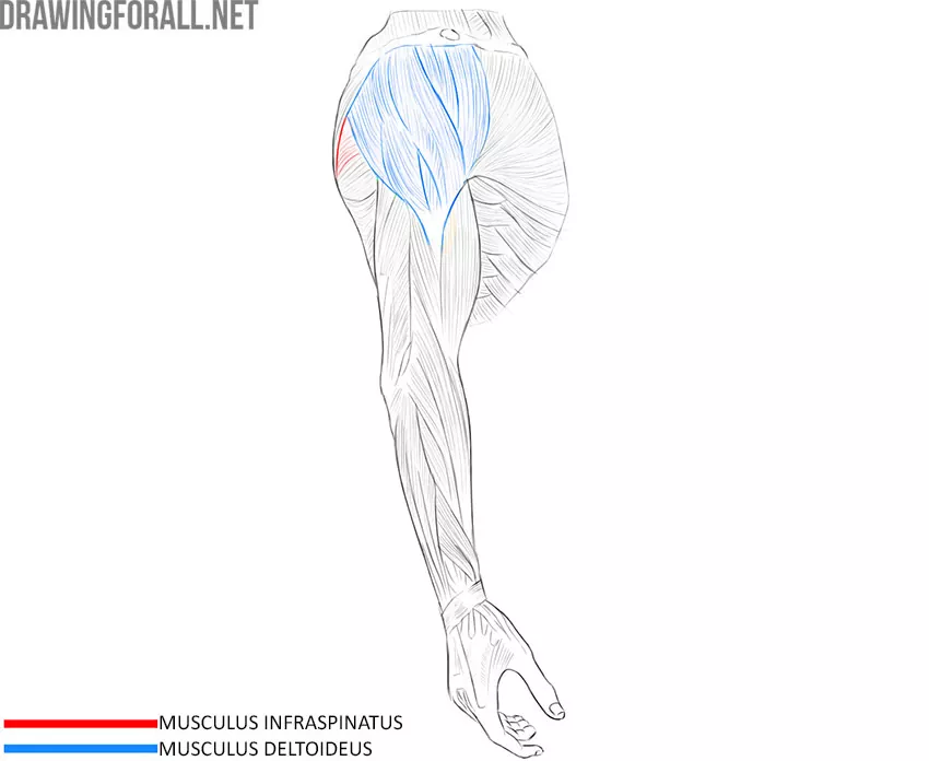
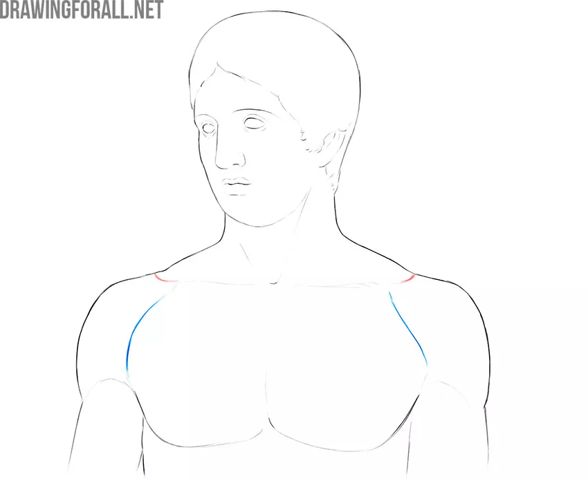
Deltoid
There are many misconceptions associated with this muscle. Many call it “shoulder” or “shoulder muscle” or “arm muscle”. In fact, the deltoid muscle refers to the muscles of the girdle of the upper limb. This muscle really forms the contour of the upper arm, especially if we are talking about a person with developed muscles. You’ve probably seen these round balls on the tops of the shoulders of comics superheroes. But despite this, the deltoid muscle is not an arm muscle.
The deltoid muscle starts from the lateral third of the clavicle, acromion, and spine of the scapula. As you can see, the deltoid muscle occupies a fairly large area. This muscle is attached to the deltoid tuberosity of the humerus. By contracting, the deltoid muscle moves the arm away from the body. The deltoid muscle is located directly under the skin, so it is very important in plastic anatomy.
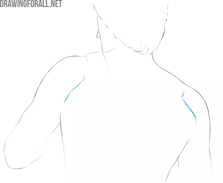
Infraspinatus Muscle
As you remember, the posterior surface of the scapula is divided into two large sections – the supraspinatus and infraspinatus fossa. The boundary between these pits is a bony protrusion called the spine of the scapula. Both the infraspinatus and the supraspinatus fossa are filled with the corresponding muscles. The infraspinatus muscle is especially large and powerful. That is why we see large parts of the infraspinatus muscle on a living person.
The infraspinatus muscle is located between the trapezius muscle and the pectoralis major muscle. This muscle is especially well visible when the arm is raised. The infraspinatus muscle begins over the entire area of the infraspinatus fossa and attaches to the upper part of the humerus. By the way, many people mistakenly consider this muscle to be one of the back muscles; this is a very popular misconception.
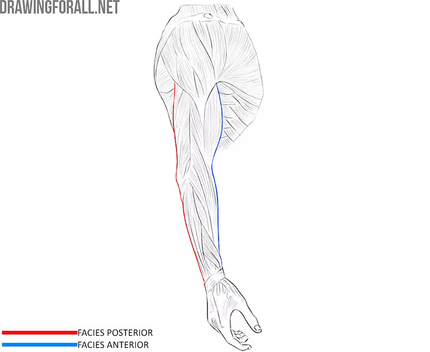
Muscles of the Shoulder
The muscles of the shoulder (as well as forearm) are classified into anterior and posterior muscles. The anterior shoulder muscles include the biceps and the brachialis. The posterior muscles include three large muscle fibers that form the powerful, large triceps and the coracobrachialis muscle.
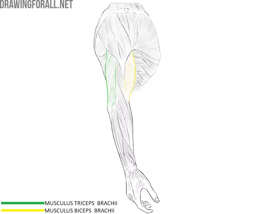
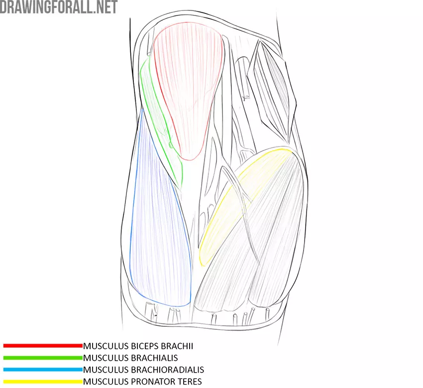
Biceps
Probably, many people know where this muscle is located, because exercises on this muscle are the most common and stereotyped in many gyms.
The biceps occupies the anterior surface of the shoulder. In athletic people, the biceps muscle forms a prominent, rounded contour, which is especially noticeable when the arm is bent. This is not surprising, because the main task of this muscle is to bend the arm at the elbow joint.
The biceps has two parts. The lateral part starts from the supra-articular tubercle of the scapula. The medial part starts from the apex of the coracoid process of the scapula. Both parts converge into a large, monolithic muscle, which spreads over the elbow joint and attaches to the tuberosity of the radius.
Triceps
The contour of the entire back of the upper arm, not covered by the deltoid muscle, is formed by the powerful triceps muscle. Interestingly, if you remove all the soft tissue on the back of the shoulder, you will only see two parts of the triceps muscle. To see the third part, you need to lift one of the two surface parts.
Only the medial triceps muscle attaches to the scapula. The other two parts start from the top of the back of the humerus. All three parts connect to form a single muscle that attaches to the olecranon of the ulna. Thus, the muscle participates in the extension of the arm in the shoulder joint and fully extends the arm in the elbow joint.
Muscles of the Forearm
This is a very numerous group of large, developed muscles. The fibers of the forearm muscles attach to the bones of the forearm and hand, which are highly mobile parts of the body. On the front and back sides of the forearm muscle are located in two layers. We will not consider the deep and thin muscles, but we will definitely study the largest muscles that form the contours of the upper limb.
Front Surface
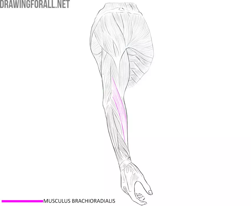
Brachioradialis muscle
This muscle is most lateral in the extended arm. As you remember, the radius corresponds to the thumb, and the ulna corresponds to the little finger. The brachioradialis muscle is also located on the side of the thumb. This muscle is especially developed in gymnasts and climbers.
Spider-Man also has very pronounced forearm muscles because he constantly hangs on the web or climbs up.
The brachioradialis muscle starts from the humerus just above the condyle and attaches to the lateral surface of the radius. The main function of the brachioradialis muscle is to flex the arm at the elbow joint. In fact, all the muscles on the front of the arm are responsible for flexion.
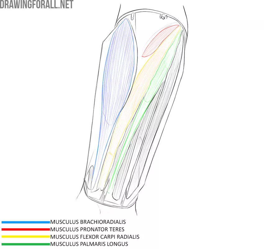
Pronator Teres Muscle
You can easily find the pronator teres using the previous muscle. The brachioradialis muscle and pronator teres form the letter Y. In people with developed muscles, this muscle is also clearly visible due to its superficial location.
The pronator teres starts from the medial epicondyle of the humerus, as well as from the coronal process of the ulna. This muscle is attached to the center of the radius from the lateral side. The pronator teres flexes the arm at the elbow and rotates the forearm.
Musculus Flexor Carpi Radialis
We continue to study the muscles of the forearm using our preparation. The musculus flexor carpi radialis is adjacent to the pronator teres. It is a long, elongated muscle that stretches from the epicondyle of the humerus to the second metacarpal bone.
As you know, each muscle goes into a dense tendon and it is the tendon that attaches to the bone. The musculus flexor carpi radialis has a long tendon that becomes visible under the skin when the wrist is flexed. The function of this muscle is to flex the hand.
Palmaris Longus Muscle
If we move a little more medially, we see another muscle with a very long tendon. It stretches from the medial epicondyle of the humerus to the hand, woven into the palmar aponeurosis – a wide connective tissue plate that is located under the skin of the palm.
The tendon of this muscle is located most superficially, so it clearly protrudes through the skin when the arm is bent. The function of this muscle is to flex the hand.
Back Surface
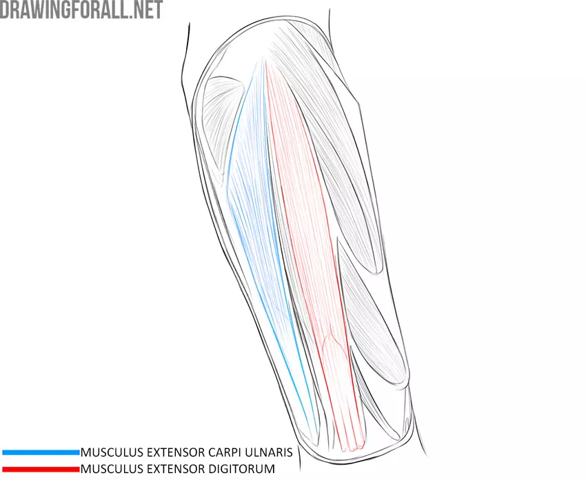
Extensor Carpi Ulnaris Muscle
If we look at the back of the forearm, we see several muscles. On a living person, two muscles are contoured, and one of them is the ulnar extensor of the wrist. As you remember, all the anterior surface muscles are flexor muscles, and all posterior surface muscles are extensor muscles.
The extensor carpi ulnaris has two parts, one of which starts from the ulna and the other from the radius. Downward, the two parts of the muscle join into one powerful muscle fiber, which passes into a long, narrow tendon and attaches to the fifth metacarpal bone. When contracting, this muscle extends the hand.
Extensor Digitorum Muscle
This is the largest and most powerful of the entire group. You can see this muscle even in people who are not professional athletes. The extensor digitorum muscle starts from the lateral epicondyle of the humerus and attaches to the distal phalanges of the fingers of the hand (except for the thumb).
During preparation, this muscle often becomes a reference point due to the large tendon, which is very visible and is located most superficially.
Muscles of the Hand
The hand is a very mobile part of the human body. The bones of the hand are controlled by two groups at once – the muscles of the forearm and the muscles of the hand. In this category, we will be looking at the muscles of the hand.
The muscles of the hand are divided into two groups – the muscles of the back of the hand and the muscles of the palm. In plastic anatomy, the muscles of the palm are important, which form the eminences of the thumb and little finger.
Muscles of the Thumb
Abductor Pollicis Brevis Muscle
This muscle is located on the lateral side of the eminence of the thumb. The abductor pollicis brevis muscle is involved in the formation of the volume of the palm because it is located directly under the skin. This muscle starts from the navicular bone and attaches to the lateral surface of the base of the proximal phalanx of the thumb.
Flexor Pollicis Brevis Muscle
This muscle is located slightly medial to the previous muscle, also directly under the skin. The short flexor of the thumb starts from the trapezoid, trapezium, and capitate bones, as well as from the base of the first metacarpal bone. The flexor pollicis brevis muscle is attached to both bones of the metacarpophalangeal joint of the thumb bone.
Muscles of the Little Finger
Palmaris Brevis Muscle
This muscle looks like a thin plate that starts from the inner edge of the palmar aponeurosis and the flexor muscle retainer and is woven into the skin of the pinky eminence.
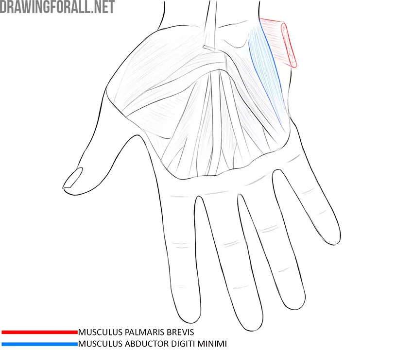
Abductor Digiti Minimi Muscle of Hand
This muscle is an excellent landmark because it occupies the most medial position of all the muscles in the palm. The abductor digiti minimi muscle of hand starts from the pisiform bone and the flexor muscle retainer and attaches to the base of the pinky phalanx.
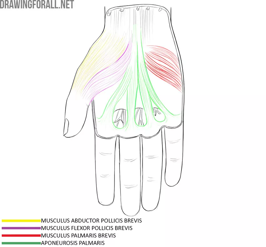
Aponeurosis palmaris
The palmar aponeurosis is a dense fiber of connective tissue that sits between the skin and muscles. The long and thick fiber of the palmar aponeurosis divides into four branches, each of which goes to the corresponding finger.
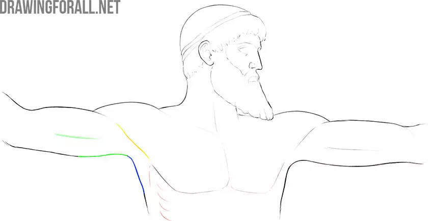
Upper Limb Topography
The upper limb is bordered by the chest, neck, and back.
The border separating the chest and upper limb is the depression between the deltoid and pectoralis major muscle. This depression is called the deltoid-thoracic sulcus.
The border separating the upper limb and the neck is a line drawn through the upper edges of the collarbones and acromion.
The border between the upper limb and the back is the posterior edge of the deltoid muscle.
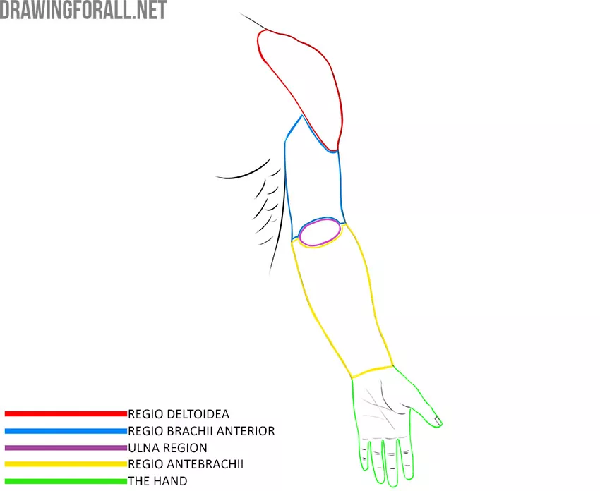
Upper Limb Regions
The boundaries of the deltoid region correspond to the boundaries of the deltoid muscle.
The upper arm region is limited from above by the lower edge of the deltoid muscle, from below – by the upper edge of the ulnar region. The upper arm region is subdivided into the anterior and posterior region. The borders of the anterior region correspond to the border of the biceps brachii, the borders of the posterior region correspond to the borders of the triceps brachii.
The ulnar region in front is represented by the ulnar fossa, behind – by the visible boundaries of the olecranon process.
The region of the forearm is bounded from above by the lower border of the cubital fossa, from below by a conditional line drawn through the styloid processes of the radius and ulna.
The hand is bounded from above by a conditional line drawn through the styloid processes of the radius and ulna.
Important Topographic Objects
Ulnar fossa
The ulnar fossa is bounded from above by the brachial muscle, medially by the circular pronator, laterally by the brachioradial muscle. The ulnar fossa becomes clearly visible when the skin and subcutaneous fat are removed, however, it can occasionally be seen on a living person.
Axillary fossa
The axillary fossa is bounded in front by the pectoralis major and minor, posteriorly by the latissimus dorsi, laterally by the serratus anterior muscle, medially by the biceps brachii and the coracohumeral muscle.