Upper Limb Skeleton Anatomy
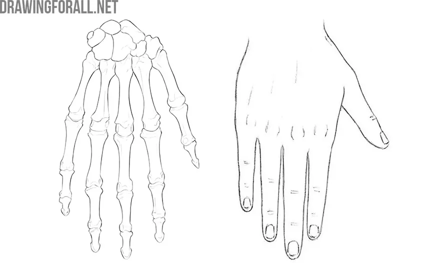
We can do some pretty remarkable things with our hands. Surgeons can perform highly precise operations on the brain, Formula 1 pit crews can change wheels in two or three seconds, and we can create new drawing tutorials for you.
We can do all these things because of our highly developed upper limbs. This is an impressive evolutionary construction that we will examine in detail here.
More precisely, we will consider the skeleton of the entire upper limb. As a rule, the upper limb, especially the hand, is a very difficult subject for artists.
The upper limb girdle is formed by only two bones – the clavicle and the scapula. It is much simpler than the pelvic girdle, isn’t it?
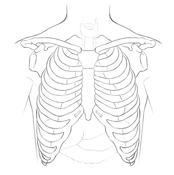
The upper limb girdle is attached to the rib cage by numerous muscles and ligaments, as well as by the sternoclavicular joint. This is what the joint looks like:
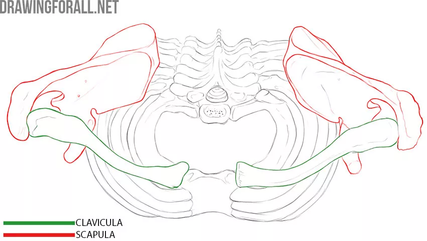
Let’s look at each bone individually.
Scapula
The scapula is a large, flat bone that lies against the back of the rib cage. It is triangular in shape. Accordingly, we can identify three angles, three borders, and several important processes on the scapula.
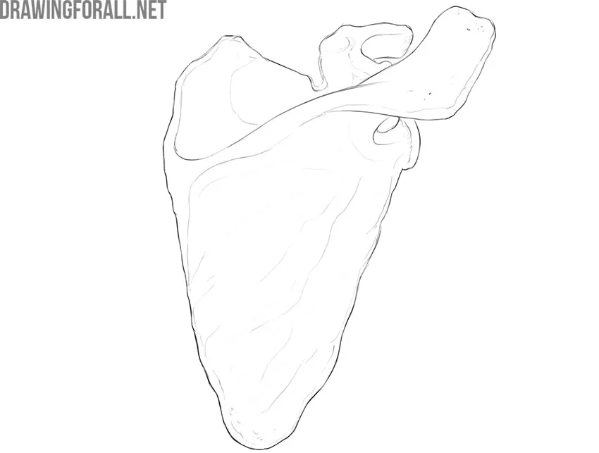
The borders of the scapula are the medial border, lateral border, and superior border. The angles have slightly different names: the superior angle, inferior angle, and lateral angle.
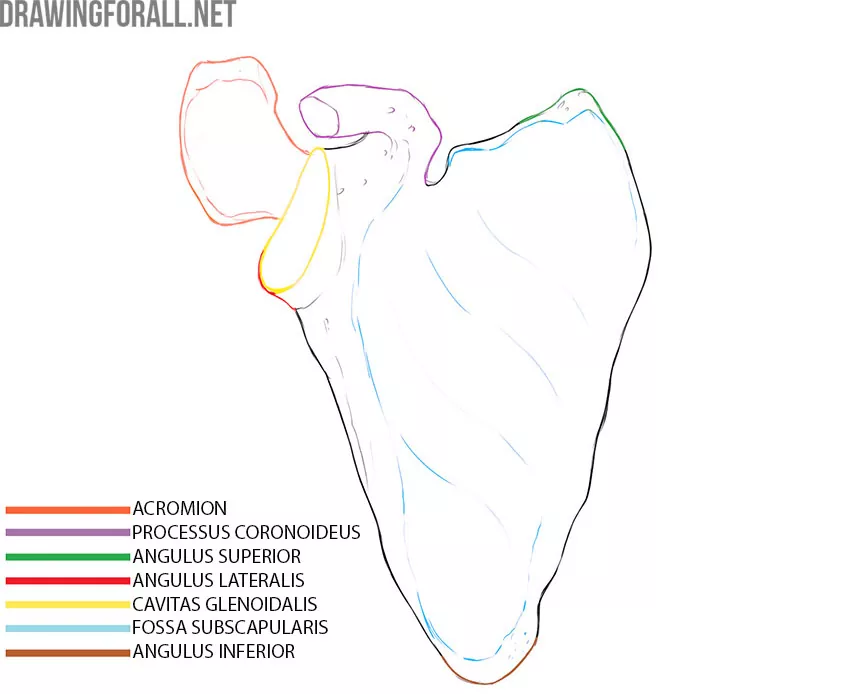
Since, as we noted, the scapula is a flat bone, it has two surfaces. One surface lies against the ribs and is called the costal surface. The opposite surface faces the back and is therefore called the dorsal surface.
On the costal surface, we can see a large fossa that occupies almost the entire area. This is the subscapular fossa (fossa subscapularis), which is filled by the fibers of the strong subscapularis muscle.
We can also see the superior (angulus superior), inferior (angulus inferior), and lateral (angulus lateralis) angles of the scapula.
If we turn the scapula over and look at the dorsal surface, we can see a large horizontal projection that runs at a slight angle.
This is the spine of the scapula (spina scapulae). The area of the dorsal surface above the spine is called the supraspinous fossa (fossa supraspinata), while the area below the spine is called the infraspinous fossa (fossa infraspinata). Both fossae are filled by muscles of the same names, which are noticeably defined in athletic people.
The spine of the scapula continues into a large, rounded projection called the acromion. The acromion forms the junction between the scapula and the clavicle. If we examine the acromion itself, we can see the acromial angle and the acromial articular surface.
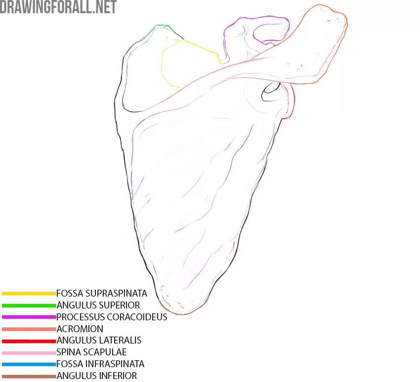
Near the acromion is the lateral angle of the scapula. At the lateral angle is an articular socket called the glenoid cavity (cavitas glenoidalis).
Just above the glenoid cavity is the coracoid process of the scapula (processus coracoideus), to which several muscles are attached.
Clavicle
The clavicle is a very important bone of the upper limb that connects the rest of the upper limb to the rib cage. It is a fairly long, curved bone.
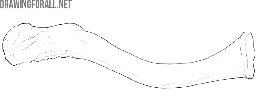
The clavicle has a shaft and two ends. One end attaches to the sternum (extremitas sternalis), while the other attaches to the acromion of the scapula (extremitas acromialis).
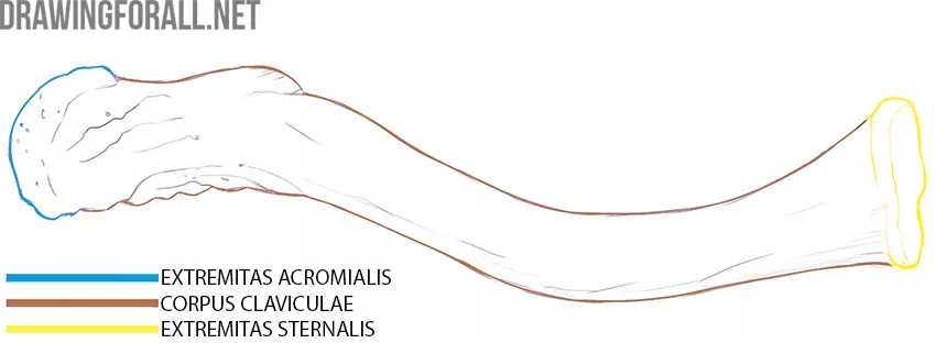
The clavicle is a bone that is clearly visible in thin people. Moreover, in a person with this type of build, the supraclavicular fossa is also clearly visible.
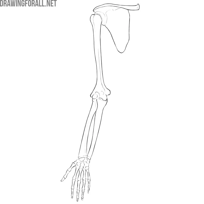
The free upper limb consists of the arm, forearm, and hand. The skeleton of the arm is formed by the humerus, while the skeleton of the forearm is formed by the ulna and radius.
The skeleton of the hand consists of many bones, which are divided into the carpal bones, metacarpal bones, and phalanges of the fingers.
Free upper limb
Humerus
The humerus is the largest bone in the upper limb. Like any other long bone, the humerus has two ends—the upper and lower ends. The ends of long bones are called epiphyses. Most of the humerus consists of its shaft, or diaphysis.
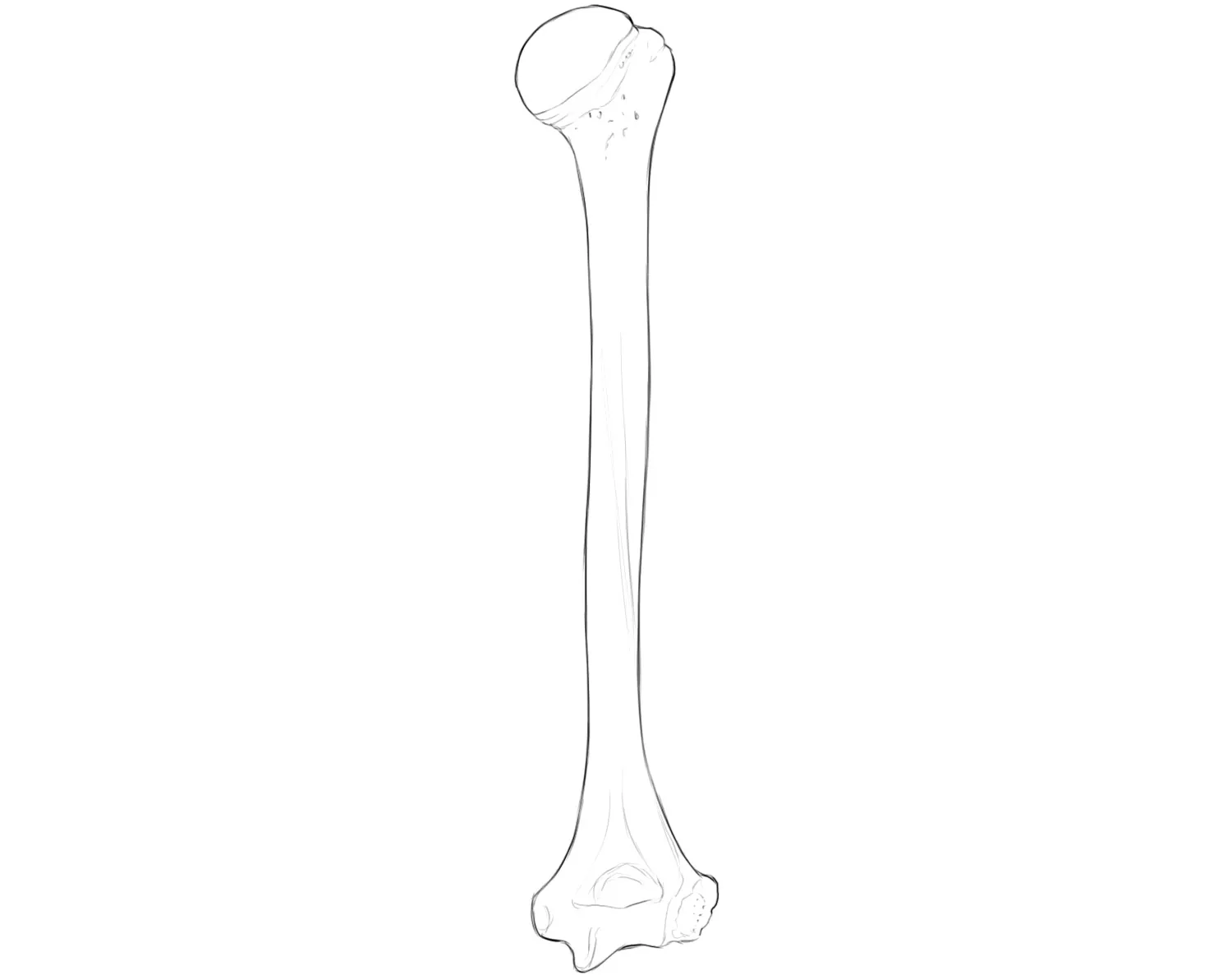
At the proximal epiphysis is the head of the humerus (caput humeri), a rounded structure that articulates with the scapula.
The head, as is common in anatomy, narrows into the anatomical neck. Moving farther toward the shaft, we encounter an even more pronounced narrowing called the surgical neck. This part has such an unusual name because it is a common site of fractures during falls.
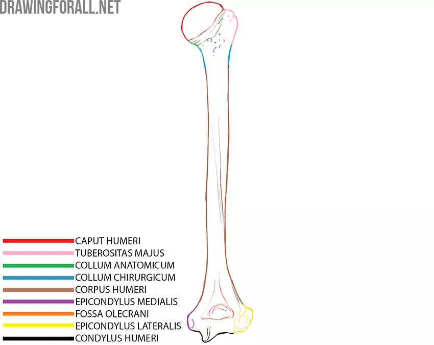
The greater tubercle and lesser tubercle are located near the head. Crests of the same names extend downward from each tubercle.
In the middle of the shaft, you can see the deltoid tuberosity, a clearly visible area with a rough texture to which the powerful deltoid muscle is attached.
At the distal epiphysis, there are small elevations above the articular surface called the epicondyles. The articular surface is called the condyle of the humerus. This surface consists of the capitulum and the trochlea of the humerus. In front of the articular surface is the coronoid fossa, while behind it is the olecranon fossa.
Radius
The humerus articulates with the ulna and radius. You may remember a simple rule: the radius is on the thumb side, and the ulna is on the little-finger side.
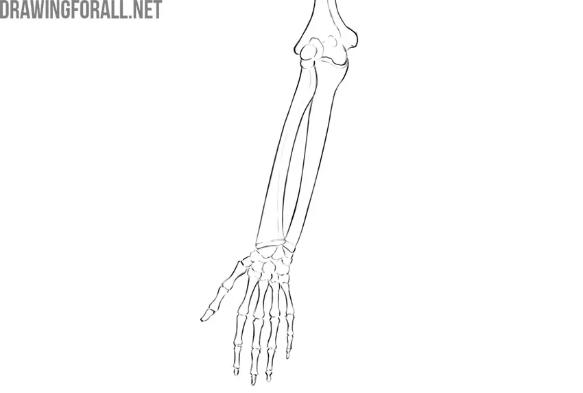
The radius is a long bone that lies between the humerus and the hand on the thumb side of the forearm. It has a head (caput radii) that continues into the neck (collum radii). A little below the neck is the radial tuberosity.
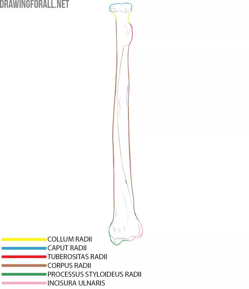
Farther down is the shaft of the radius, which ends in an expanded distal end. On the lateral side, we can see the styloid process of the radius (processus styloideus radii), while on the opposite side is the ulnar notch (incisura ulnaris) for articulation with the ulna. The diagram marks the location of the ulnar notch on the posterior surface.
Ulna
The ulna is the second bone that forms the forearm. Like all long bones, it has two ends and a shaft. The proximal end of the ulna articulates with the humerus and radius. The distal end of the ulna articulates with the radius and the bones of the hand.
The ulna has a distinctive landmark that makes it easy to identify and distinguish from the radius, even if you have forgotten the little-finger and thumb rule. This landmark is the large olecranon at the proximal end.
At the proximal end, you can also see the coronoid process, which is smaller. Just below these processes, on the shaft of the bone, is the ulnar tuberosity.
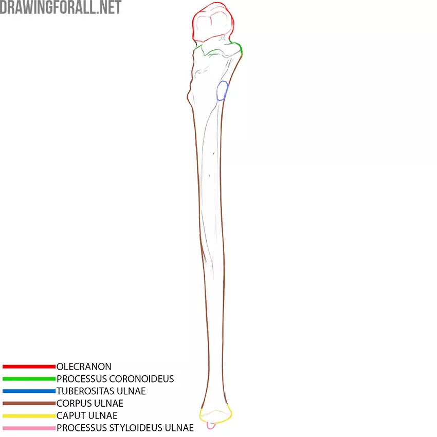
The shaft of the ulna resembles a triangular prism because of the interosseous border projecting toward the lateral side.
At the distal end of the ulna, we can see the head of the ulna (caput ulnae), which articulates with the radius, and the styloid process.
Hand bones
The bones of the hand are numerous bones that form one of the most mobile parts of the human body. The joints between these bones allow us to make delicate and precise movements that are unattainable for other living beings on our planet.
The bones of the hand are divided into the carpal bones, metacarpal bones, and phalanges of the fingers.
The wrist contains eight small, dense bones arranged in two rows: proximal and distal. The proximal row includes the scaphoid, lunate, triquetrum, and pisiform bones. The distal row includes the trapezium, trapezoid, capitate, and hamate bones.
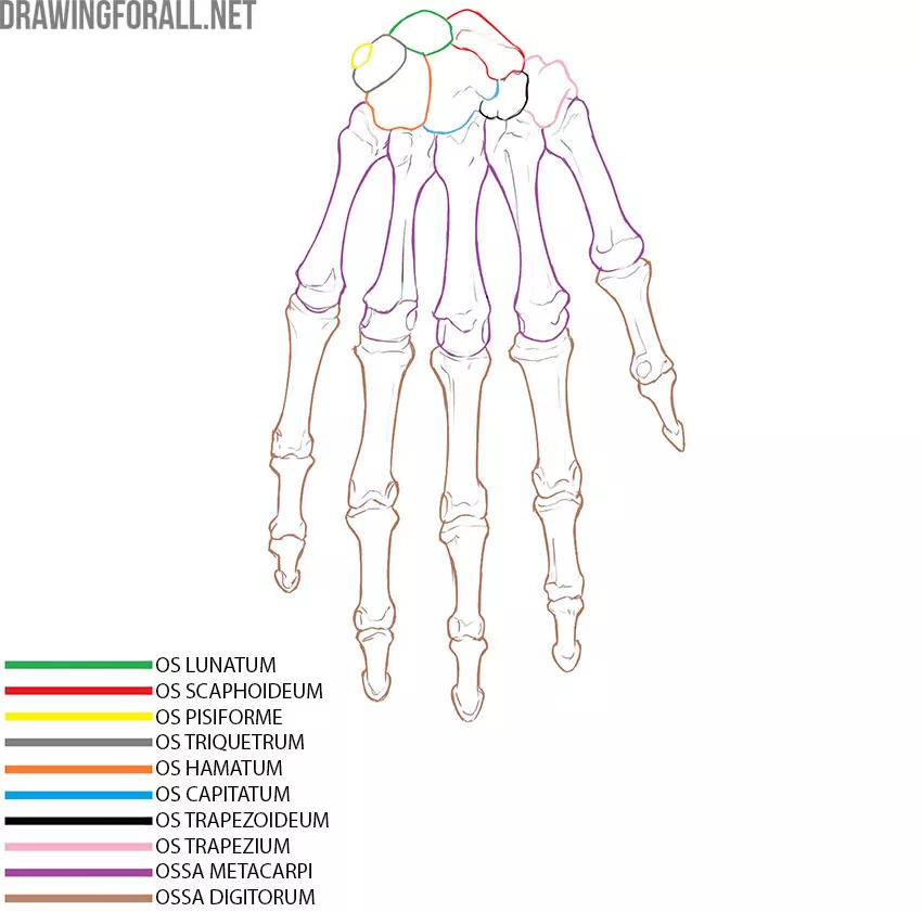
The metacarpals are long bones that differ greatly from the bones of the wrist. The metacarpal bones do not have individual names; they are simply numbered from first to fifth, beginning with the thumb and ending with the little finger.
Thus, the metacarpal bone of the thumb is the first metacarpal, while the metacarpal bone of the little finger is the fifth metacarpal.
The most distal bones of the hand are the finger bones. These are short, tubular bones also called phalanges. The thumb has two phalanges: proximal and distal. The remaining fingers have three phalanges: proximal, middle, and distal.