Lower Limbs Muscle Anatomy
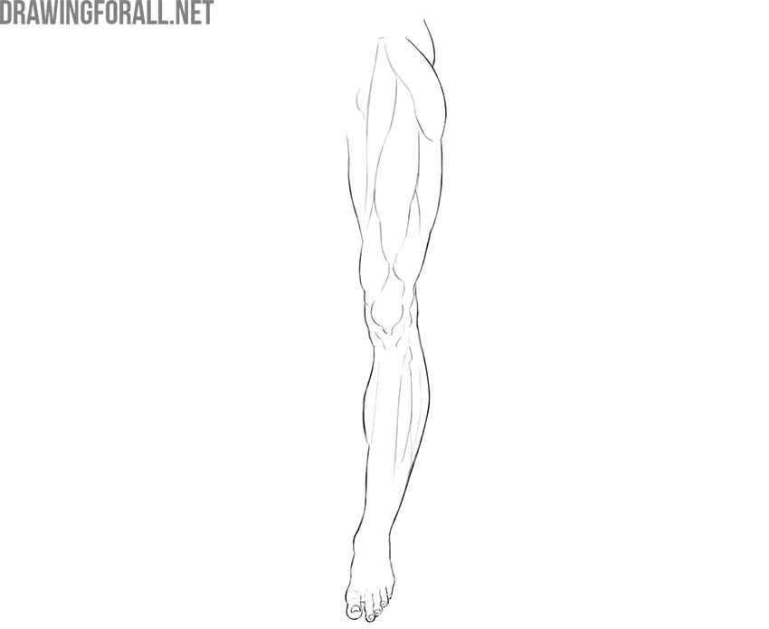
In this guide, we will tell you about the anatomy of the lower limbs. This is a very important topic for artists because the position of the legs shapes the pose and figure of the character.
Muscles of the Girdle of the Lower Limbs
The muscles of the lower limb girdle connect the bones of the lower limb girdle and the free lower limb into a single mechanism. These muscles are divided into superficial and deep. The deep ones are not of fundamental importance for plastic anatomy, but we will definitely analyze some superficial ones.
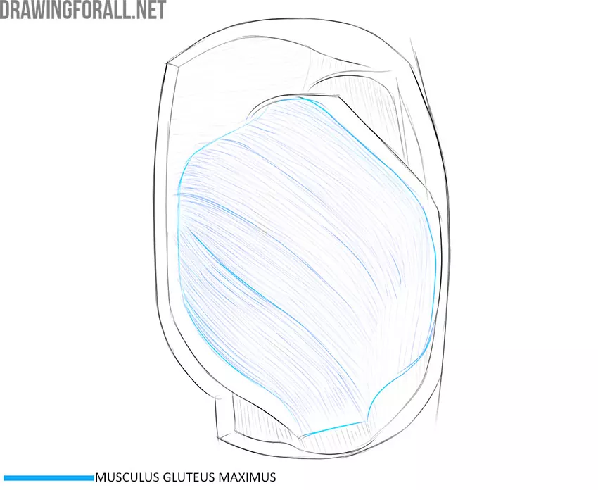
Gluteus Maximus Muscle
This rounded, raised muscle forms the contour of the back of the pelvis. This muscle is especially developed in powerlifters and gymnasts. The gluteus maximus is the most superficial muscle of all the pelvic muscles and is located just under the skin and adipose tissue.
The gluteus maximus muscle begins from the back sides of all the bones of the pelvic girdle and from the tendon of the spinal erectors. The attachment point of this muscle is the gluteal tuberosity of the femur and the iliotibial tract.
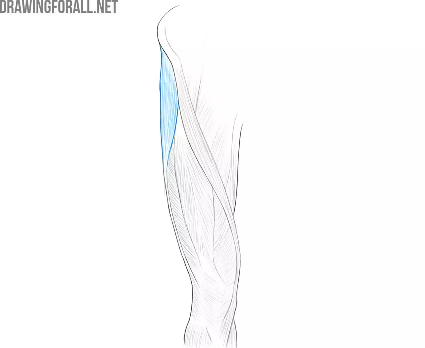
Tensor Fasciae Latae Muscle
This muscle occupies the most lateral position of all muscles studied today. We can see a small muscle bundle that merges into a powerful tendon structure called fascia lata (iliotibial tract).
The fascia lata tensor begins from a small area on the wing of the ilium just below the superior anterior iliac spine. Going down, it is woven into the iliotibial tract, which reaches the knee joint.
Thigh Muscles
The muscles of the thigh are very strong and massive because these muscles take on a significant part of the load associated with upright posture. The thigh muscles are divided into the muscles of the front, back, and medial groups.
Front Group
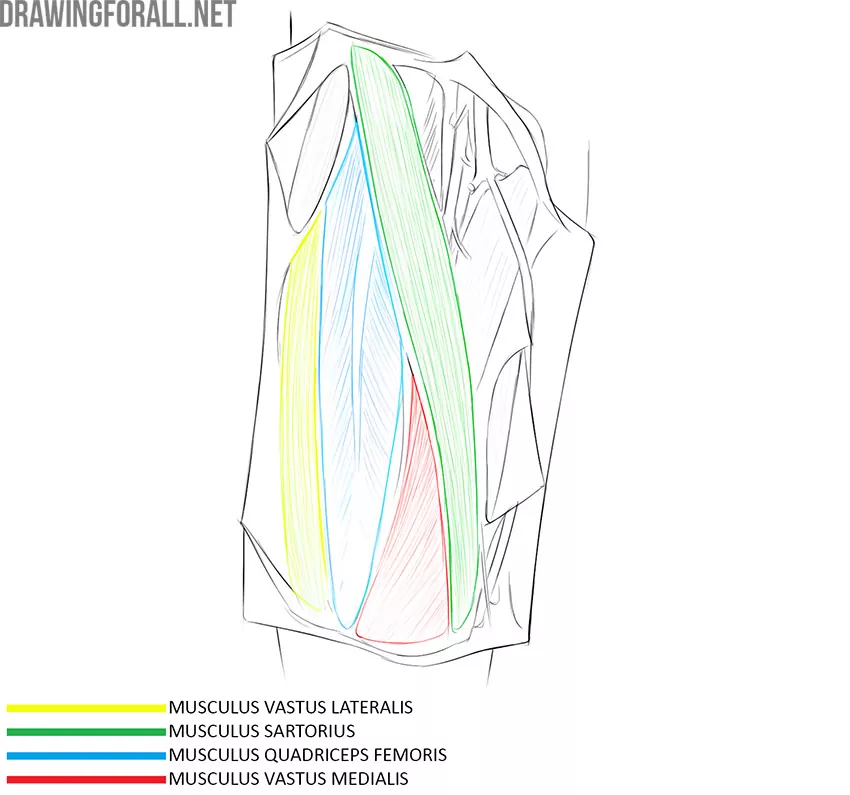
Quadriceps Femoris Muscle
It is the largest of the thigh muscles. As you can see from the name, the quadriceps muscle is consists of four parts. Each part of the quadriceps muscle has its own name.
The constituent parts of the quadriceps muscle of the thigh start from different places, but, going downwards, these parts connect into a powerful tendon that spreads over the knee joint and attaches to the tuberosity of the tibia. The patella is located in the tissue of this tendon.
Rectus Femoris
This muscle occupies the entire front of the thigh. Rectus femoris muscle begins with an elongated, thin tendon from the spina iliaca anterior superior (pelvis). Then the tendon passes into a wide, powerful muscle layer, which narrows again near the knee joint and is woven into the common tendon of the quadriceps femoris muscle.
Vastus Medialis
This part of the quadriceps muscle is located medial to the rectus femoris. A small area of the vastus medialis muscle is covered by the rectus femur muscle. From below, the fibers of the vastus medialis muscle are woven into the common tendon of the quadriceps muscle.
Vastus Lateralis
This part of the quadriceps muscle is located lateral to the rectus muscle and occupies almost the entire anterolateral surface of the thigh. The lateral muscle is covered in front by the rectus femoris muscle. Also, this muscle is bordered by the fascia lata (iliotibial tract). The vastus lateralis starts from the trochanter major of the femur and from the intertrochanteric line. Like all other parts of the quadriceps muscle, the vastus lateralis muscle passes into the common tendon and attaches to the tibial tuberosity.
Back Group
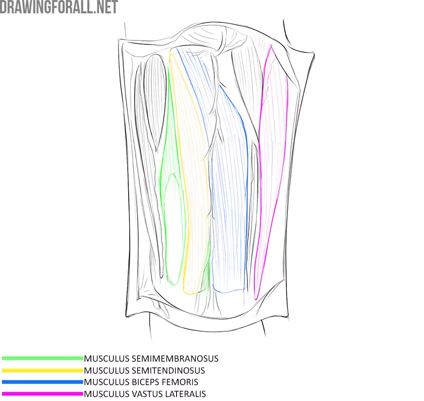
Biceps Femoris Muscle
The biceps femoris has a long and short part. The short part is located under the long part, so it is irrelevant in plastic anatomy. The long part covers the most lateral position on the back surface. This part starts from the ischial tuberosity and attaches to the caput of the fibula.
Semitendinosus Muscle
This muscle is a long, elongated strap that runs along the medial edge of the back of the thigh. The semitendinosus muscle is located directly under the skin and partially overlaps the semimembranosus muscle. This muscle starts from the ischial tuberosity and attaches to the tibial tuberosity.
The semitendinosus and biceps muscles form a well-marked arch-like figure. This is especially noticeable if the person stands stretched out to his full height.
The semimembranosus muscle is located directly below the semitendinosus muscle.
Shin Muscles
The shin is the part of the leg that is located between the knee joint and the foot. The bone base of the shin is made up of the fibula and tibia. The shin muscles control the movements of the foot and toes, so this is a fairly large group.
We will not study all the shin muscles, because among them there are very deep muscles that are not important for plastic anatomy. We will only study the superficial muscles that form the contours of the shin.
Anterior Calf Muscle Group
These muscles are located on the front surface of the shin and extend the foot and toes.
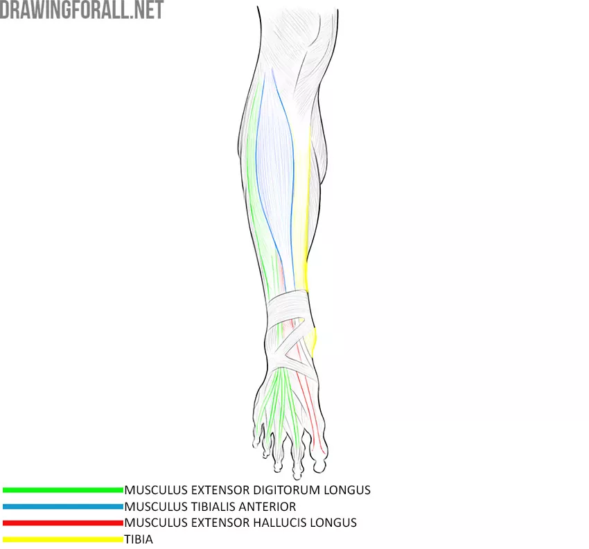
Tibialis Anterior Muscle
It is a very large and powerful muscle. The anterior tibial muscle forms the anterior rounded contour of the lower leg. This muscle fills the space lateral to the anterior edge of the tibia.
The anterior tibial muscle starts from the lateral condyle and from the upper half of the lateral surface of the tibia. Downward, this muscle passes into a long thin tendon, which attaches to the plantar surface of the medial sphenoid bone and the base of the 1st metatarsal bone.
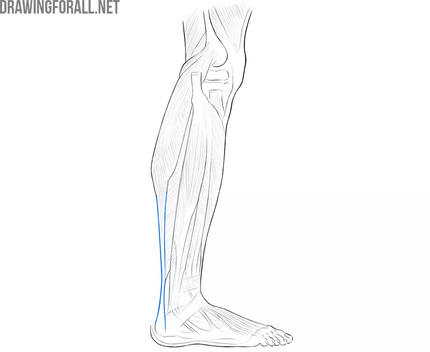
Extensor Digitorum Longus Muscle
This muscle is adjacent to the tibialis anterior muscle from the medial side. The extensor digitorum longus is a slightly thinner muscle that is located throughout the lower leg. Downward, the extensor digitorum longus passes into four long, thin tendons that diverge along the corresponding fingers and attach to the distal phalanges.
The tendons of the extensor digitorum longus are clearly contoured under the skin when the muscle contracts.
Extensor digitorum longus muscle begins from the lateral condyle of the tibia and the anterior surface of the body of the fibula.
Extensor Hallucis Longus Muscle
We cannot see the main part of this muscle due to the fact that it is almost completely overlapped by the anterior tibial muscle. However, the extensor hallucis longus is very noticeable under the skin, especially during times of muscle tension.
The extensor hallucis longus starts from the medial surface of the lower 2/3 of the fibula and attaches to the distal phalanx of the big toe.
Back Muscle Group of the Shin
This group includes the most powerful and most visible shin muscles. Back muscles of the shin are located in several layers, and we will get acquainted with the largest and most superficial of them.
Triceps Surae Muscle
This is the only muscle of the superficial layer. However, it is a very large and wide muscle with several parts. In addition, it forms the entire posterior and partially anterior contours of the lower leg. The triceps muscle consists of three parts, and each part has its own name. We will consider only two parts that are more superficial.
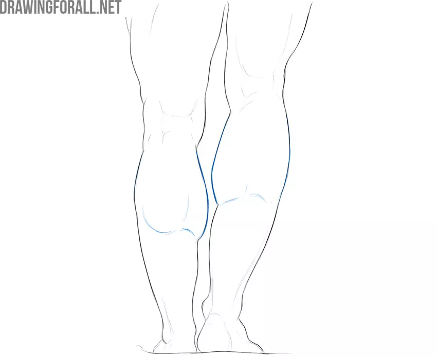
Gastrocnemius Muscle
This is the common name for two large, massive muscles that are located on the back of the lower leg. Distinguish between the medial and lateral parts of the gastrocnemius muscle. If the gastrocnemius muscle is sufficiently developed, we see the graceful contours of the lower leg with a pronounced narrowing towards the foot.
The main function of the gastrocnemius muscle is to flex the foot. The work of this muscle is most noticeable when you look at professional ballet dancers in those moments when they are standing on their toes.
The lateral head of the gastrocnemius muscle starts from the outer surface of the lower epiphysis of the femur. The medial head starts from the popliteal surface above the medial condyle of the femur.
Under the lateral and medial parts of the triceps muscle of the lower leg is a flat, broad muscle called the soleus muscle. The soleus muscle is not visible unless the medial and lateral heads are removed, so we will not dwell on this muscle in detail.
Achilles Tendon
Achilles tendon is a tendon into which all three heads of the triceps muscle of the lower leg pass. It attaches to the heel and is therefore also called the heel tendon. The history of the name of this tendon is connected with the history of the Trojan War, more precisely, with the myth about one of its alleged participants. We are talking about Achilles – one of the main warriors of the Greeks who (according to the legends of Homer) besieged Troy.
Achilles’ mother, the goddess Thetis, bathed her little son in the waters of the Styx River. This gave the future hero complete invulnerability. However, while bathing, Thetis held Achilles by the heel. It was in this heel, in the midst of one of the last battles before the fall of Troy, Achilles was mortally wounded by the Trojan prince Paris.
In honor of this ancient myth, the heel tendon is called the Achilles tendon.
Muscles of the Foot
The muscles of the foot are classified into the muscles of the sole of the foot and muscles of the dorsum of the foot. We will study only those muscles that are located superficially since it is these muscles that are important in plastic anatomy.
Muscles of the Rear of the Foot
The muscles on the rear of the foot are responsible for the extension of the toes. These muscles are located under the tendons of the anterior leg muscles and, in fact, duplicate their function.
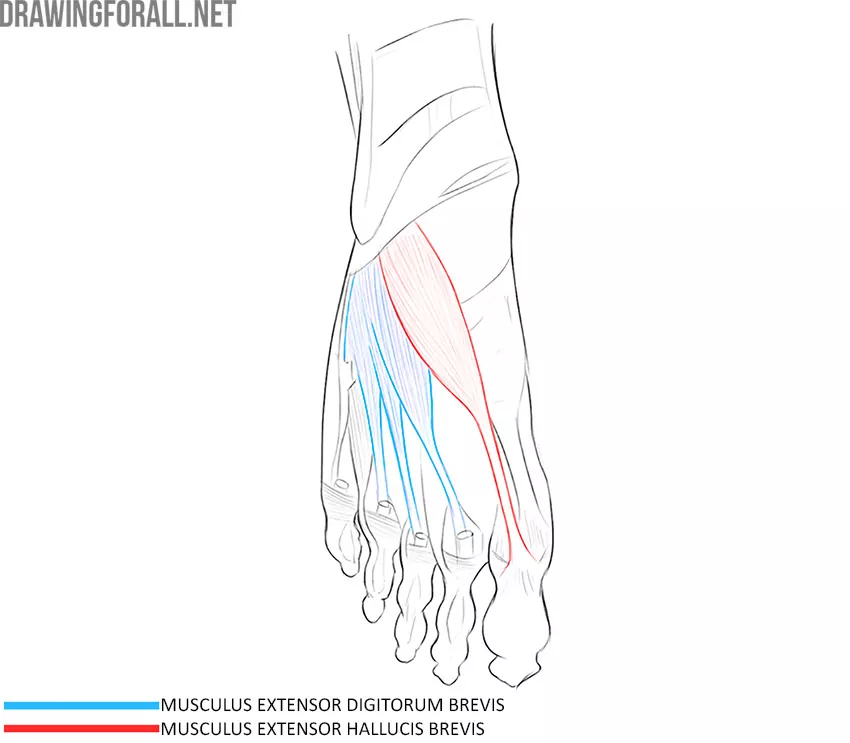
Extensor Digitorum Brevis Muscle
It is a fairly large, broad muscle that divides into four sections. Each of the parts is directed to the corresponding finger, becomes a long, thin tendon, and attaches to the proximal phalanx of the finger. This muscle starts from the anterior surface of the heel bone.
Extensor Hallucis Brevis Muscle
There are usually separate muscles for the thumb. In this case, we also see a special muscle that controls the extension of the big toe. The extensor hallucis brevis starts from the superior surface of the calcaneus and attaches to the proximal phalanx of the big toe.
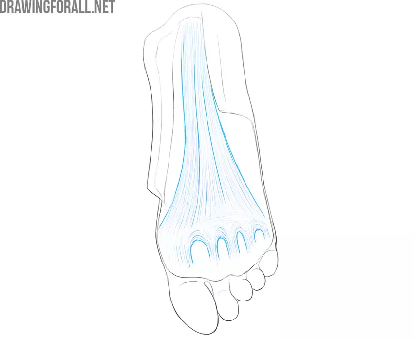
Plantar Aponeurosis
Almost all muscles of this group are located under the plantar aponeurosis. It is a very strong layer of connective tissue that sits under the skin and is divided into bundles corresponding to the fingers.
The plantar aponeurosis is a dense lamina of connective tissue that sits directly under the skin of the sole. The plantar aponeurosis consists of three large bundles of connective tissue, which are perpendicularly connected to the transverse fascia of the sole. Five connective tissue bundles extend from this connection to the corresponding fingers.
When extending the fingers or standing on toes, the plantar aponeurosis clearly appears under the skin.
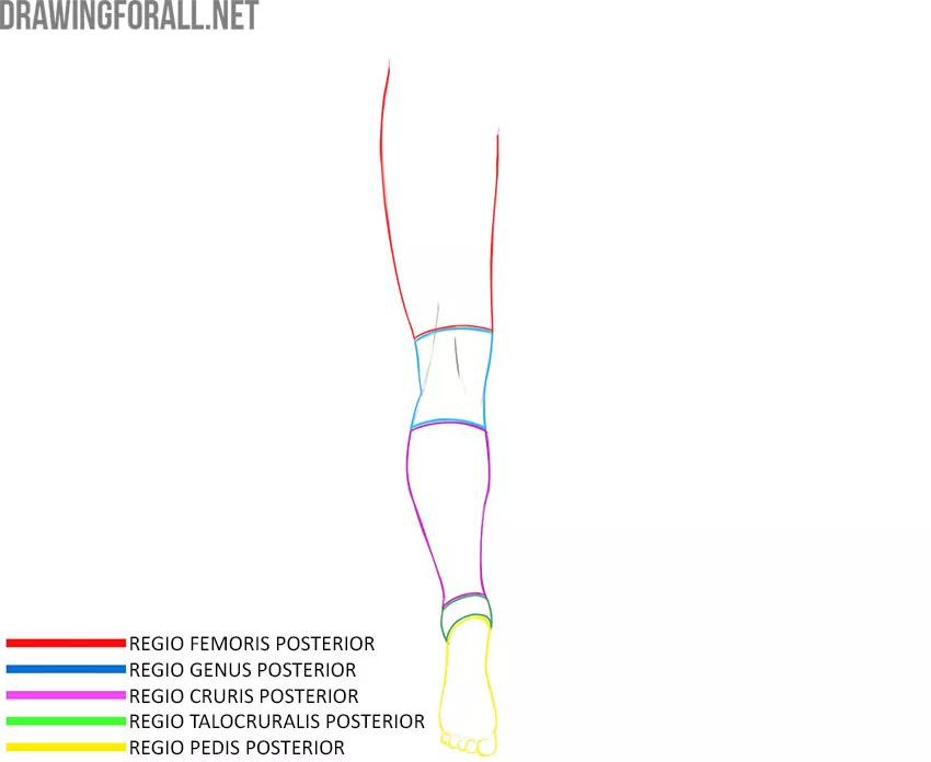
Lower Limb Topography
The lower limb is bordered by the abdomen and back. The border separating the lower limb and the abdomen is the inguinal ligament. The border separating the back and lower limb is a line drawn through the iliac crest and the apex of the coccyx.
Lower Limb Regions
Gluteal Region
The gluteal region corresponds to the gluteus maximus muscle.
Thigh Region
The thigh area has front and back surfaces. The front surface is bounded from above by the inguinal ligament, from below by a horizontal line drawn 3-5 centimeters above the patella.
The posterior surface is bounded from above by the gluteal fold, and from below by a horizontal line drawn 3-5 centimeters above the projection of the patella onto the posterior surface.
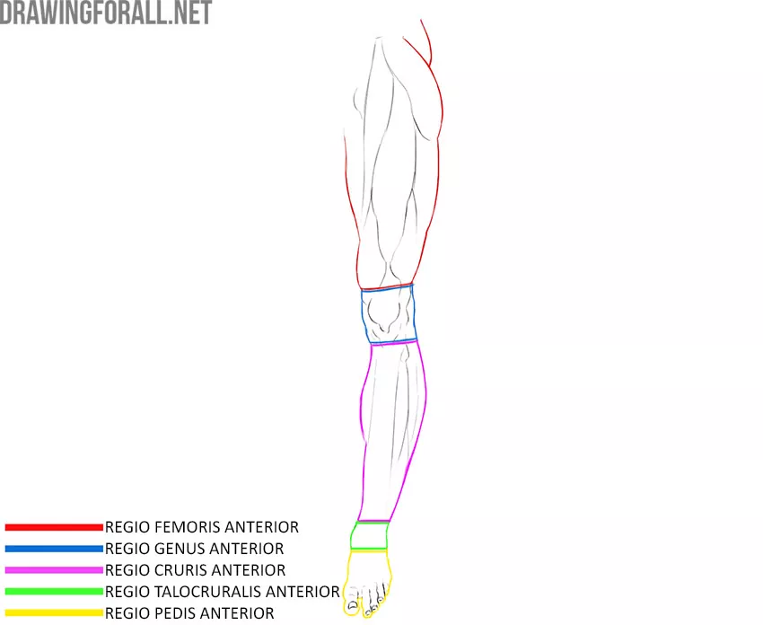
Knee Region
The knee region also has front and back surfaces.
The front area of the knee is bounded from above by a horizontal line drawn 3-5 centimeters above the patella, and from below by a horizontal line drawn through the tuberosity of the tibia. The posterior region of the knee corresponds to the boundaries of the popliteal fossa.
The popliteal fossa is diamond-shaped. The popliteal fossa is bounded from above laterally by the biceps femoris, from above medially by the semimembranosus muscle, below by the lateral and medial parts of the gastrocnemius muscle.
Shin Region
The front region of the lower leg is bounded from above by a horizontal line drawn through the tuberosity of the tibia, and from below by a line drawn through the bases of both ankles.
The posterior region of the lower leg is bounded from above by an annular line drawn through the tuberosity of the tibia, and from below by a line drawn through the bases of both ankles.
Ankle Region
This area corresponds to the location of the ankle joint.
Foot Region
The region of the foot is bounded at the top by a line connecting the lower edges of both ankles.