Torso Muscles Anatomy

The muscles of the torso shape a person’s appearance in many ways. Muscles of the torso, as well as muscles in the arms or legs, can give the impression of a thin or athletic person.
But, above all, these muscles are of great physiological importance. The muscles of the back and abdomen help to keep the spine in an upright position and serve as reliable protection for the abdominal organs.
The muscles of the chest help to make various movements with the arms, and these muscles also participate in breathing, setting the chest in motion.
We analyze plastic anatomy, that is, the structure of those anatomical structures that form the contour of the body. Therefore, today we will show you the superficial muscles of the back, chest, and abdomen.
Pectoral Muscles
The pectoral muscles are divided into superficial and deep. The superficial pectoral muscles attach to the bones of the upper limb. These muscles move the arm. The deep muscles are located between the ribs and move the ribcage, helping with breathing.
Pectoralis Major
The most prominent pectoral muscle is the pectoralis major. This superficial muscle is located just under the skin, so it is very visible in athletic people. Slightly higher and lateral to the pectoralis major muscle is the deltoid muscle of the shoulder. Below are the abdominal muscles and the serratus anterior muscle.
The pectoralis major muscle starts from the medial part of the clavicle, the anterior surface of the sternum, the upper costal cartilages, and the aponeurosis of the external oblique muscle.
The pectoralis major muscle attaches to the lateral lip of the intertubercular groove of the humerus. Remember, the superficial pectoral muscles are involved in movements of the upper limb.

Serratus Anterior Muscle
The long parts of this muscle cover the ribs. It is a fairly powerful muscle that sits superficially, so you can see the serratus anterior muscle in athletic people with a small amount of subcutaneous fat.
The most important feature that distinguishes the serratus anterior muscle from many other muscles associated with the ribs is the location of the muscle parts just above the ribs, and not between them.
The serratus anterior muscle begins on the outer surfaces of the first eight or nine ribs and attaches along the medial border of the scapula.

This group also includes the pectoralis minor muscle and the subclavius muscle, which we can only see if the pectoralis major muscle is removed, so these muscles are not particularly important for plastic anatomy.
Muscles of the Back
The muscles of the back are a very large group of muscles, the main tasks of which are to control the movements of the spine and maintain the spine in an upright position. The back muscles in humans are strongly developed due to upright posture. The muscles of the back lie in several layers, and we will only examine those muscles that form the contour of the body.
Trapezius Muscle
This is a paired muscle, and if we consider only the left or only the right trapezius muscle, we will see a triangular muscle shape. If we look at the contours of both muscles together, then we really see a figure that looks like a trapezoid.
The trapezius muscle is located under the skin. It is noticeable when you strain your back, raise your shoulders, and tilt your head back slightly. In this position, this muscle becomes visible even in people with a normal physique.
The trapezius muscle starts from the occipital bone of the skull and from the spinous processes of the cervical and thoracic vertebrae. This is a fairly broad muscle. It attaches to the lateral part of the clavicle, the acromion, and the spine of the scapula.
Latissimus Dorsi Muscle
This is another powerful and large superficial paired back muscle. The latissimus dorsi muscles cover much of the lower back like a corset. In athletic people, this muscle can be clearly visible even from the front.
These areas are sometimes referred to as wings. A developed latissimus dorsi muscle in combination with the deltoid and pectoralis major muscles can create a figure with a very wide upper body and a narrow pelvis.
The powerful latissimus dorsi muscle begins from the lower thoracic vertebrae, thoracolumbar fascia, iliac crest, and lower ribs. The latissimus dorsi attaches to the intertubercular groove of the humerus. It extends, adducts, and medially rotates the arm. The latissimus dorsi is well known to people who work out because it is strongly engaged during pull-ups.

Erector Spinae Muscles
These muscles occupy a deep position, that is, they are covered by the skin, fascia, and other muscles. However, the erector spinae form a powerful and large muscle group, so their contours can be visible on a living person.
The erector spinae muscles extend along the vertebral column and have numerous attachments to the pelvis, sacrum, ribs, vertebrae, and skull.
The erector spinae muscles hold the vertebral column in an upright position and extend it. They also help extend and laterally flex the head and spine.

Abdominal Muscles
The abdominal muscles are divided into anterior, lateral, and posterior groups. For plastic anatomy, the muscles that form the anterior and lateral walls are of particular importance.
Rectus Abdominis Muscle
This muscle looks like a long, wide strap that extends vertically along the front of the abdomen. Throughout its entire length, the rectus abdominis muscle is crossed by several tendinous intersections. This is how the characteristic rectangular areas commonly called abs are formed.
The rectus abdominis muscle starts from the pubic crest and pubic symphysis and attaches to the xiphoid process and the costal cartilages of the fifth, sixth, and seventh ribs.
The main function of the rectus abdominis is flexion of the spinal column, which is the opposite action to extension by the erector spinae muscles. It also helps compress the abdominal contents.

Abdominal External Oblique Muscle
The fibers of this muscle form part of a muscular corset stretched between the ribs and the pelvis. The external oblique muscle of the abdomen starts from the outer surfaces of the lower eight ribs, with its upper attachments interdigitating with the serratus anterior muscle. This further emphasizes the visible relief of the serratus anterior muscle.
The external oblique muscle attaches to the linea alba, pubic tubercle, and anterior part of the iliac crest. Its lower aponeurotic edge forms the inguinal ligament, which extends from the anterior superior iliac spine to the pubic tubercle.
The abdominal external oblique muscle, like the rectus muscle, is clearly visible in people with a low amount of subcutaneous fat or with very developed musculature.

In this illustration, we can see the rectus abdominis muscle against the background of the deeper abdominal muscles:

Chest Topography
Chest Borders
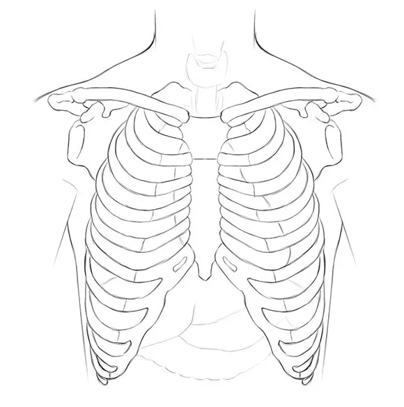
The border that separates the chest from the neck runs from the jugular notch of the sternum along the superior edges of the clavicles and the acromioclavicular joints. Further, the line passes to the back and continues to the spinous process of the 7th cervical vertebra.
The lower border of the chest is projected onto a line that goes from the xiphoid process of the sternum along the costal arch to the spinous process of the 12th thoracic vertebra.
The lateral border of the chest includes the deltopectoral groove, which separates the chest from the free upper limb.
Chest Regions
In topographic anatomy, the chest has anterior and posterior walls. Many anatomists do not consider the back as a separate region, dividing it into the posterior wall of the chest and the posterior wall of the abdomen.
Presternal Region
The boundaries of the presternal region coincide with the boundaries of the sternum.
Pectoral Region
In athletic people, the boundaries of the pectoral region coincide with the boundaries of the pectoralis major muscle. More reliable landmarks for the pectoral area are:
- The upper edge of the clavicle – the upper border;
- The lower edge of the 6th rib – the lower border;
- The lateral edge of the sternum – the medial border;
- Deltopectoral groove – lateral border.
Substernal Region
The substernal region is bounded:
- Above – by the 6th rib (in athletic people, this border coincides with the contour of the pectoralis major muscle);
- Below – by the costal arch;
- Medially – by the lateral edge of the sternum;
- Laterally – by the middle axillary line.
Scapular Region
The scapular region has the following boundaries:
- Above – coincides with the upper border of the chest (see above);
- Below – a horizontal line drawn through the lower angle of the scapula;
- Medially – a line drawn through the medial edge of the scapula;
- Laterally – the posterior edge of the deltoid muscle and the middle axillary line.
Infrascapular Region
The infrascapular region has the following boundaries:
- Above – a line drawn through the lower angle of the scapula;
- Below – the 12th rib;
- Medially – the paravertebral line;
- Laterally – the middle axillary line.
Vertebral Region
The vertebral region has the following boundaries:
- Above – coincides with the upper border of the chest (see above);
- Below – a horizontal line drawn through the spinous process of the 12th thoracic vertebra;
- Laterally – the paravertebral lines.

Abdominal Regions
Like the chest, the abdomen has front and back walls, with the back wall corresponding to the lower half of the back.
The abdomen has the following borders.
Above, the abdomen is bounded by the lower edges of the 10th pair of ribs. Below, the abdomen is bordered by the lesser pelvis. The boundary between the abdomen and the lesser pelvis is the terminal line, which we cannot see on a living person.
The anterior abdominal wall is divided into three areas – epigastrium, mesogastrium, and hypogastrium. To draw the boundaries of these areas, we need to draw two conventional horizontal lines.
The upper line is drawn through the lowest points of the 10th pair of ribs, and the lower one through the anterior superior iliac spines. Thus, the upper region is the epigastrium, the middle is the mesogastrium, and the lower is the hypogastrium.
The back wall of the abdomen is formed by the muscles of the lower back and the portion of the spine that extends into the lumbar region. The upper border of the lumbar region is the lower edge of the 12th rib, and the lower border of the lumbar region is the iliac crest.