Neck Muscles Anatomy
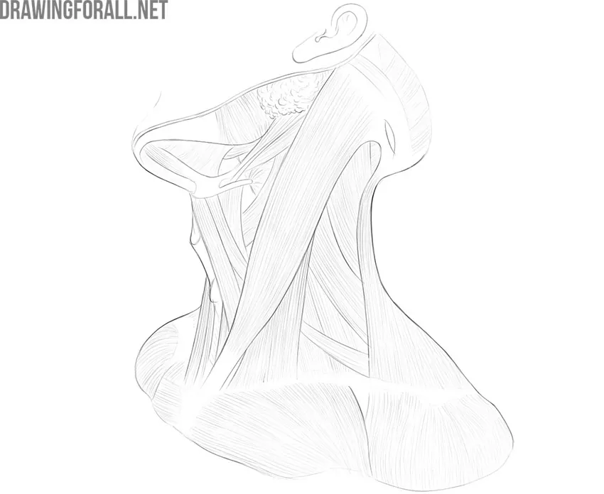
As you know, the neck is the part of the body that sits between the head and torso. The neck has no external bone protective structures, so it is quite mobile. Neck mobility is necessary primarily to rotate the head and keep the head upright. This is a rather important task that is performed not only by the bones but also by the muscles of the neck. We will talk about them.
The neck muscles are divided into three large groups, which correspond to the depth of their location:
- Neck muscles of the superficial layer;
- Neck muscles of the middle layer;
- Deep neck muscles.
For plastic anatomy, the muscles of the neck are of particular importance, which shapes the appearance of this part of the body and is noticeable during contraction. These are, first of all, the superficial muscles and some muscles of the middle layer. In this article, we are going to study these muscles.
The Muscles of the Superficial Layer
Platysma Muscle
It is a very wide flat muscle of the neck that covers the entire front of the neck like a collar. The platysma subcutaneous muscle of the neck (platysma) extends from the chin to the pectoral region. Many modern anatomists refer this muscle to the facial muscles of the head because some of its fibers are woven into the skin in the area of the base of the body of the lower jaw. Therefore, under tension, the subcutaneous muscle of the neck forms a characteristic facial expression with tense face skin and downward corners of the mouth.
The muscle begins from the superficial fascia of the chest slightly below the clavicle.
The Sternocleidomastoid Muscle
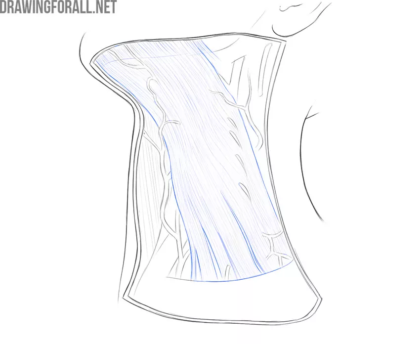
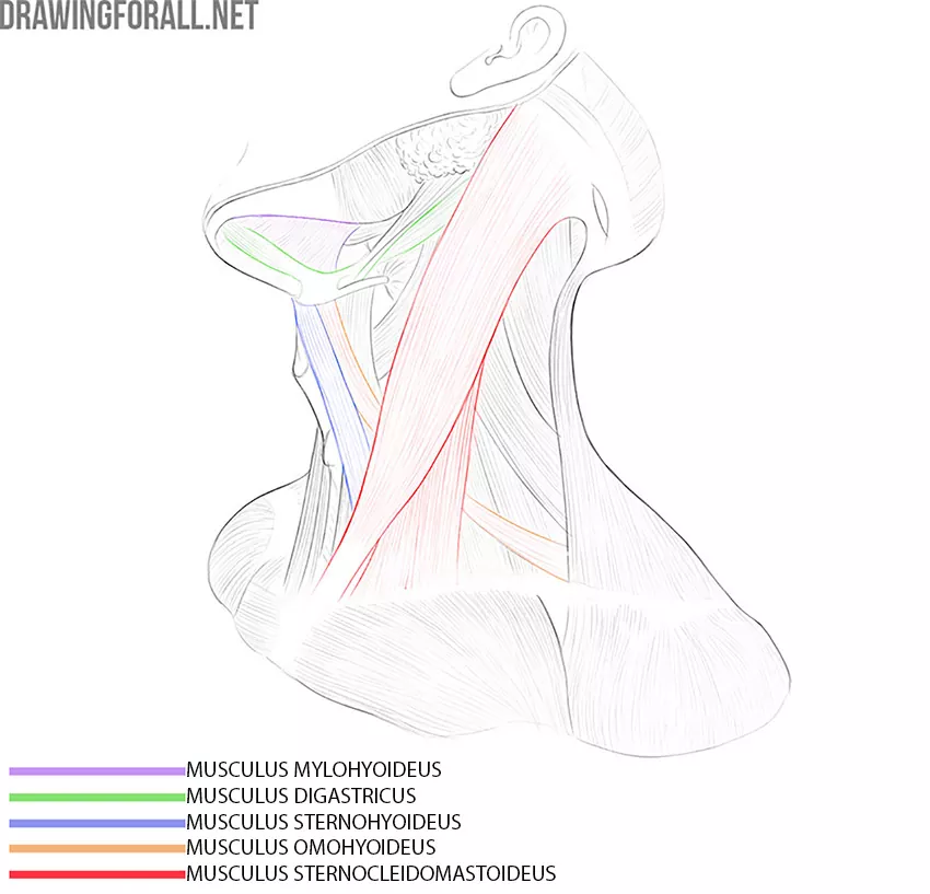
This muscle also belongs to the superficial, although it is very different from the thin and wide subcutaneous muscle. The sternocleidomastoid muscle is a large, rather powerful paired bundle that starts from the sternum and clavicle and attaches to the mastoid process of the skull. This muscle is located directly under the platysma.
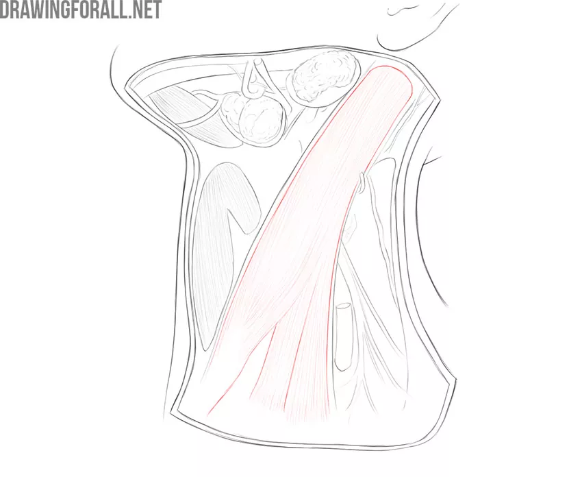
In a person without excess body weight or diseases that deform the contour of the neck, this muscle visible very well through the skin when turning the head.
The Muscles of the Middle Layer
The muscles of the middle layer are also divided into two groups according to their location. The first group is located above the hyoid bone, and the second is below the hyoid bone. We will dwell in detail only on some of the muscles of this group, which are important for plastic anatomy.
Digastric Muscle
It is a rather unusual muscle that looks like an obtuse angle connecting the anterior edge of the mandible and the area between the mastoid and styloid processes of the skull. In fact, it is just two parts, the anterior of which starts from the digastric fossa of the lower jaw, and the second from the mastoid notch. Both parts are attached to the hyoid bone.
Mylohyoid Muscle
The mylohyoid muscle looks like a wide plate, which is located just behind the digastric muscle. This muscle starts from the maxillary-hyoid line of the lower jaw and attaches to the hyoid bone. Like all muscles in this group, the mylohyoid muscle raises the hyoid bone. Also, the mylohyoid muscle is involved in lowering the lower jaw.
Muscles Located Below the Hyoid Bone
These muscles are longer and more elongated compared to the muscles that are located below the hyoid bone. The muscles located below the hyoid bone include the omohyoid muscle, sternohyoid muscle, sternothyroid muscle, and thyrohyoid muscle.
We will analyze two muscles that are most important for topography and plastic anatomy.
Omohyoid Muscle
It is a long muscle fiber that runs from the hyoid bone to the upper edge of the scapula. The omohyoid muscle divides the neck into several areas in the form of triangles. This is a very good reference point for studying other anatomical formations of the neck.
Sternohyoid Muscle
It is the most prominent of the three remaining muscles of the middle layer below the hyoid bone. The sternohyoid muscle is located parallel to the trachea and connects the manubrium sternum to the hyoid bone. The function of this muscle, like the rest of the muscles of the entire group, is to lower the hyoid bone.
Neck Borders
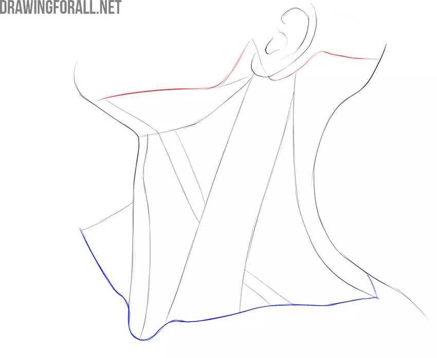
The upper border of the neck is a line drawn from the edge of the lower jaw to the mastoid process, then along the superior nuchal line of the occipital bone to the external occipital protuberance.
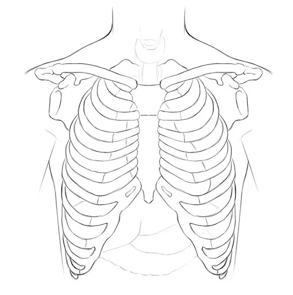
The lower border of the neck is drawn from the manubrium of the sternum to the upper edges of the clavicles, then along the acromions to the spinous process of the 7th cervical vertebra.
Neck Regions
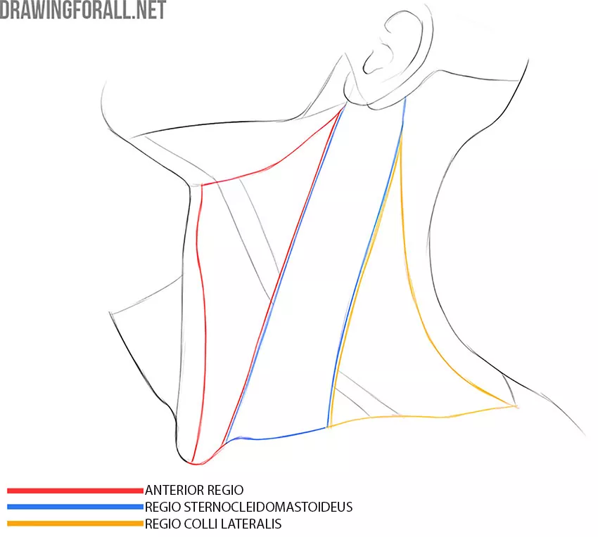
The anterior region of the neck is bordered from above by the upper edge of the lower jaw, from below by the handle of the sternum, and laterally by the anterior edges of the sternocleidomastoid muscles. The anterior region is an unpaired region.
The lateral region of the neck is paired, each area is bordered in front by the rear margin of the sternocleidomastoid muscle, behind by the anterior edge of the trapezius muscle, below by the clavicle. The upper border is absent since the trapezius and sternocleidomastoid muscles form an acute angle.
The sternocleidomastoid region corresponds to the sternocleidomastoid muscle.
The posterior region is bounded from above by the superior nuchal line of the occiput, from below by a conditional line drawn through the acromions of the scapula and the spinous process of the 7th cervical vertebra, laterally by the lateral edges of the trapezius muscles. The back of the neck is an unpaired area.
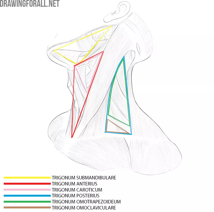
Triangles of the Neck
The submandibular triangle is bounded by both parts of the digastric muscle and the lower edge of the lower jaw.
The anterior triangle of the neck is bounded by the anterior edges of the sternocleidomastoid muscles and the lower edge of the lower jaw.
The posterior triangle is bounded by the rear margin of the sternocleidomastoid muscle, the anterior edge of the trapezius muscle and the clavicle
The carotid triangle is bounded by the anterior edge of the sternocleidomastoid muscle, the posterior part of the digastric muscle and the posterior edge of the omohyoid muscle.
The omoclavicular triangle is bounded by the clavicle, the omohyoid muscle and the posterior edge of the sternocleidomastoid muscle.
The omotrapezoid triangle is bounded by the trapezius muscle, the sternocleidomastoid muscle, and the omohyoid muscle.